The MYL9 Antibody (CAB3039) is a high-quality antibody developed for reliable detection and analysis of target proteins. This antibody, produced in rabbits, is highly specific to human MYL9 and has been validated for use in Western blot applications. By binding to the MYL9 protein, this antibody allows for accurate detection and analysis in a variety of cell types, making it ideal for investigations in cell biology and cancer research.MYL9, also known as myosin regulatory light chain 9, is essential for the regulation of smooth muscle contraction and cell motility. Dysfunction of MYL9 has been linked to various diseases, including cancer and cardiovascular disorders, making it a promising target for therapeutic intervention.
This antibody is validated for use in WB, ELISA, IF-P applications and has demonstrated reactivity against Human, Mouse, Rat samples.
Product Name:
MYL9 Antibody
SKU:
CAB3039
Size:
20μL, 100μL
Reactivity:
Human, Mouse, Rat
Conjugate:
Unconjugated
Immunogen:
Synthetic peptide. This information is considered to be commercially sensitive.
Recommended starting concentration is 1 μg/mL. Please optimize the concentration based on your specific assay requirements.
Synonyms:
LC20, MLC2, MRLC1, MYRL2, MLC-2C, MMIHS4, MYL9
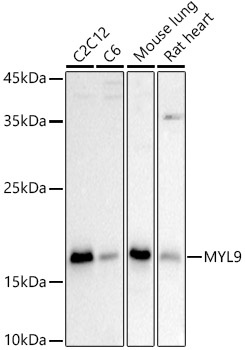
Positive Sample:
C2C12, C6, Mouse lung, Rat heart
Cellular Localization:
Cell Cortex, Cytoplasm, Cytosol, Myofibril, Z Disc.
Calculated MW:
20kDa
Observed MW:
18kDa
Myosin, a structural component of muscle, consists of two heavy chains and four light chains. The protein encoded by this gene is a myosin light chain that may regulate muscle contraction by modulating the ATPase activity of myosin heads. The encoded protein binds calcium and is activated by myosin light chain kinase. Two transcript variants encoding different isoforms have been found for this gene.
Purification Method
Affinity purification
Gene ID
10398
RRID
AB_2764844
Buffer Information
Store at -20℃. Avoid freeze / thaw cycles. Buffer: PBS containing 50% glycerol, preserved with proclin300 or sodium azide, pH 7.3.
Western blot analysis of various lysates using MYL9 Rabbit pAb (CAB3039) at 1:1000 dilution. Secondary antibody: HRP-conjugated Goat anti-Rabbit IgG (H+L) (CABS014) at 1:10000 dilution. Lysates/proteins: 25μg per lane. Blocking buffer: 3% nonfat dry milk in TBST. Detection: ECL Basic Kit (AbGn00020). Exposure time: 90s.
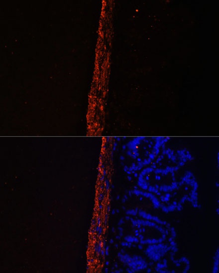
Immunofluorescence analysis of Rat intestine using MYL9 Rabbit pAb (CAB3039) at dilution of 1:100. Blue: DAPI for nuclear staining.
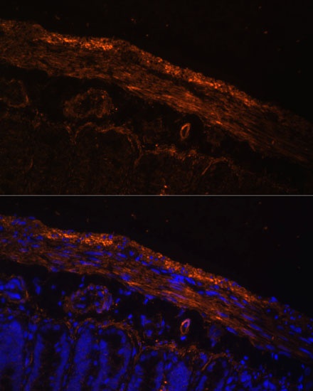
Immunofluorescence analysis of Mouse intestine using MYL9 Rabbit pAb (CAB3039) at dilution of 1:100. Blue: DAPI for nuclear staining.