The Phospho-NDRG1-T346 Antibody (CABP0792) is a high-quality antibody developed for reliable detection and analysis of target proteins. This antibody, produced in rabbits, is highly specific to the phosphorylated form of NDRG1 at threonine 346 and is validated for use in Western blot applications. By targeting this phosphorylation site, researchers can gain insights into the signaling pathways and functions regulated by NDRG1 in different cell types.NDRG1, also known as N-Myc downstream-regulated gene 1, is a key player in tumor suppression and has been implicated in the progression of various cancer types.
This antibody is validated for use in WB, ELISA applications and has demonstrated reactivity against Human, Mouse, Rat samples.
Product Name:
Phospho-NDRG1-T346 Antibody
SKU:
CABP0792
Size:
20μL, 100μL
Reactivity:
Human, Mouse, Rat
Conjugate:
Unconjugated
Immunogen:
Synthetic peptide. This information is considered to be commercially sensitive.
Sequence:
SHTS E
Tested Applications:
WBELISA
Recommended Dilution:
WB
1:500 - 1:1000
ELISA
Recommended starting concentration is 1 μg/mL. Please optimize the concentration based on your specific assay requirements.
This gene is a member of the N-myc downregulated gene family which belongs to the alpha/beta hydrolase superfamily. The protein encoded by this gene is a cytoplasmic protein involved in stress responses, hormone responses, cell growth, and differentiation. The encoded protein is necessary for p53-mediated caspase activation and apoptosis. Mutations in this gene are a cause of Charcot-Marie-Tooth disease type 4D, and expression of this gene may be a prognostic indicator for several types of cancer. Alternatively spliced transcript variants encoding multiple isoforms have been observed for this gene.
Purification Method
Affinity purification
Gene ID
10397
RRID
AB_2771356
Buffer Information
Store at -20℃. Avoid freeze / thaw cycles. Buffer: PBS with 0.01% thimerosal,50% glycerol,pH7.3.
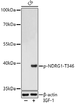
Western blot analysis of lysates from C6 cells, using Phospho-NDRG1-T346 Rabbit pAb (CABP0792) at 1:1000 dilution. C6 cells were treated with IGF-1 (50 ng/ml) at 37℃ for 30 minutes after serum-starvation overnight. Secondary antibody: HRP-conjugated Goat anti-Rabbit IgG (H+L) (CABS014) at 1:10000 dilution. Lysates/proteins: 25μg per lane. Blocking buffer: 3% nonfat dry milk in TBST. Detection: ECL Basic Kit (AbGn00020). Exposure time: 180s.