The Phospho-p38 MAPK-T180 Polyclonal Antibody (CABP1434) is a high-quality antibody developed for reliable detection and analysis of target proteins. This antibody, derived from rabbits, is highly specific to phosphorylated p38 MAPK at threonine 180 in human samples and has been validated for use in Western blotting applications.The phosphorylation of p38 MAPK at T180 is a critical event in the activation of this signaling pathway, leading to downstream effects on gene expression and cellular responses.
This antibody is validated for use in WB, IHC-P, ELISA applications and has demonstrated reactivity against Human, Mouse, Rat samples.
Product Name:
Phospho-p38 MAPK-T180 Polyclonal Antibody
SKU:
CABP1434
Size:
20μL, 100μL
Reactivity:
Human, Mouse, Rat
Conjugate:
Unconjugated
Immunogen:
Synthetic peptide. This information is considered to be commercially sensitive.
Sequence:
TGYV A
Tested Applications:
WBIHC-PELISA
Recommended Dilution:
WB
1:500 - 1:1000
IHC-P
1:50 - 1:200
ELISA
Recommended starting concentration is 1 μg/mL. Please optimize the concentration based on your specific assay requirements.
NIH/3T3 treated with UV, C6 treated with Anisomycin
Cellular Localization:
Cytoplasm, Nucleus.
Calculated MW:
41kDa
Observed MW:
42kDa
The protein encoded by this gene is a member of the MAP kinase family. MAP kinases act as an integration point for multiple biochemical signals, and are involved in a wide variety of cellular processes such as proliferation, differentiation, transcription regulation and development. This kinase is activated by various environmental stresses and proinflammatory cytokines. The activation requires its phosphorylation by MAP kinase kinases (MKKs), or its autophosphorylation triggered by the interaction of MAP3K7IP1/TAB1 protein with this kinase. The substrates of this kinase include transcription regulator ATF2, MEF2C, and MAX, cell cycle regulator CDC25B, and tumor suppressor p53, which suggest the roles of this kinase in stress related transcription and cell cycle regulation, as well as in genotoxic stress response. Four alternatively spliced transcript variants of this gene encoding distinct isoforms have been reported.
Purification Method
Affinity purification
Gene ID
1432
Buffer Information
Store at -20℃. Avoid freeze / thaw cycles. Buffer: PBS containing 50% glycerol, preserved with proclin300 or sodium azide, pH 7.3.
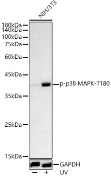
Western blot analysis of lysates from NIH/3T3 cells, using Phospho-p38 MAPK-T180 Rabbit pAb (CABP1434) at 1:500 dilution. NIH/3T3 cells were treated with UV at room temperature for 15-30 minutes. Secondary antibody: HRP-conjugated Goat anti-Rabbit IgG (H+L) (CABS014) at 1:10000 dilution. Lysates/proteins: 25μg per lane. Blocking buffer: 3% nonfat dry milk in TBST. Detection: ECL Basic Kit (AbGn00020). Exposure time: 60s.
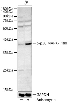
Western blot analysis of lysates from C6 cells, using Phospho-p38 MAPK-T180 Rabbit pAb (CABP1434) at 1:500 dilution. C6 cells were treated with Anisomycin (25 μg/mL) at 37℃ for 20 minutes. Secondary antibody: HRP-conjugated Goat anti-Rabbit IgG (H+L) (CABS014) at 1:10000 dilution. Lysates/proteins: 25μg per lane. Blocking buffer: 3% nonfat dry milk in TBST. Detection: ECL Basic Kit (AbGn00020). Exposure time: 180s.
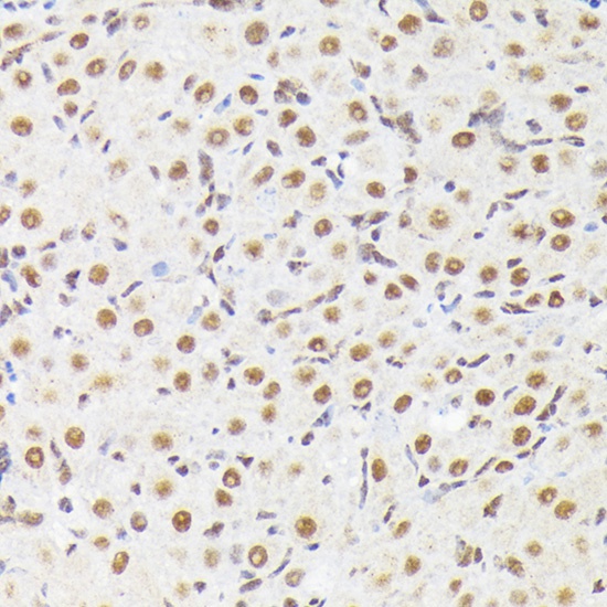
Immunohistochemistry analysis of paraffin-embedded Rat ovary using Phospho-p38 MAPK-Y182 Rabbit pAb (CABP1434) at dilution of 1:50 (40x lens). High pressure antigen retrieval performed with 0.01M Citrate buffer (pH 6.0) prior to IHC staining.
 at 1:500 dilution. NIH/3T3 cells were treated by UV at room temperature for 15-30 minutes. Secondary antibody: HRP Goat Anti-Rabbit IgG (H+L) at 1:10000 dilution. Lysates/proteins: 25μg per lane. Blocking buffer: 3% nonfat dry milk in TBST.")
 at 1:500 dilution. NIH/3T3 cells were treated by UV at room temperature for 15-30 minutes. Secondary antibody: HRP Goat Anti-Rabbit IgG (H+L) at 1:10000 dilution. Lysates/proteins: 25μg per lane. Blocking buffer: 3% nonfat dry milk in TBST.")


