The Phospho-p70 S6 Kinase 1-T389 Antibody (CABP0564) is a high-quality antibody developed for reliable detection and analysis of target proteins. This antibody, produced in rabbits, shows high reactivity with human samples and is validated for use in Western blotting applications.P70S6K1 is a serine/threonine kinase that is involved in multiple cellular processes, including cell proliferation, survival, and metabolism. Phosphorylation of p70S6K1 at threonine 389 is a crucial event that activates its kinase activity and promotes downstream signaling pathways. This antibody specifically recognizes the phosphorylated form of p70S6K1 at threonine 389, enabling researchers to investigate the regulation and function of this important signaling molecule.
This antibody is validated for use in WB, IF/ICC, ELISA applications and has demonstrated reactivity against Human, Mouse, Rat samples.
Product Name:
Phospho-p70 S6 Kinase 1-T389 Antibody
SKU:
CABP0564
Size:
20μL, 100μL
Reactivity:
Human, Mouse, Rat
Conjugate:
Unconjugated
Immunogen:
Synthetic peptide. This information is considered to be commercially sensitive.
Sequence:
GFTY VA
Tested Applications:
WBIF/ICCELISA
Recommended Dilution:
WB
1:500 - 1:5000
IF/ICC
1:50 - 1:200
ELISA
Recommended starting concentration is 1 μg/mL. Please optimize the concentration based on your specific assay requirements.
This gene encodes a member of the ribosomal S6 kinase family of serine/threonine kinases. The encoded protein responds to mTOR (mammalian target of rapamycin) signaling to promote protein synthesis, cell growth, and cell proliferation. Activity of this gene has been associated with human cancer. Alternatively spliced transcript variants have been observed. The use of alternative translation start sites results in isoforms with longer or shorter N-termini which may differ in their subcellular localizations. There are two pseudogenes for this gene on chromosome 17.
Purification Method
Affinity purification
Gene ID
6198
RRID
AB_2771385
Buffer Information
Store at -20℃. Avoid freeze / thaw cycles. Buffer: PBS containing 50% glycerol, preserved with proclin300 or sodium azide, pH 7.3.
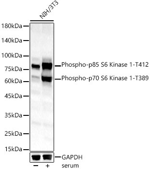
Western blot analysis of lysates from NIH/3T3 cells, using Phospho-p70 S6 Kinase 1-T389 Rabbit pAb (CABP0564) at 1:1000 dilution. NIH/3T3 cells were treated with 10% FBS at 37℃ for 30 minutes after serum-starvation overnight. Secondary antibody: HRP-conjugated Goat anti-Rabbit IgG (H+L) (CABS014) at 1:10000 dilution. Lysates/proteins: 25μg per lane. Blocking buffer: 3% nonfat dry milk in TBST. Detection: ECL Basic Kit (AbGn00020). Exposure time: 180s.
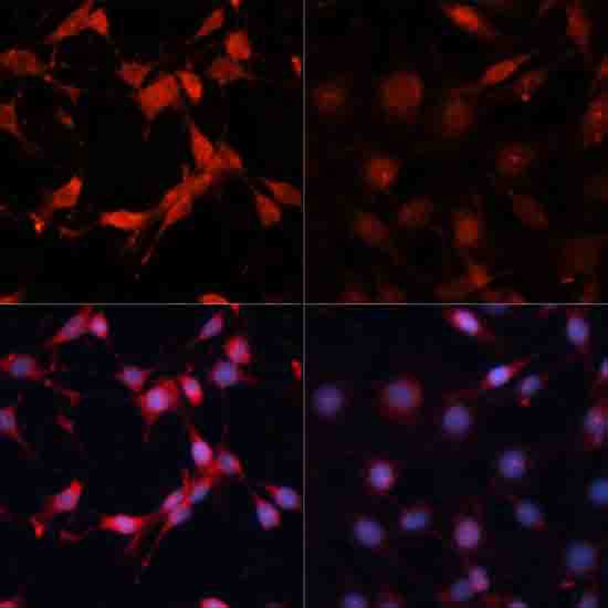
Immunofluorescence analysis of C6 cells using Phospho-p70 S6 Kinase 1-T389 Rabbit pAb (CABP0564) at dilution of 1:100. C6 cells were treated with Serum-starvation overnight at 37℃.