The Phospho-PDGFRA-Y1018 Antibody (CABP0615) is a high-quality antibody developed for reliable detection and analysis of target proteins. This antibody, raised in rabbits, is highly specific and reactive with human samples, making it ideal for use in Western blot applications. By targeting the phosphorylated tyrosine 1018 residue of the PDGFRA protein, this antibody enables precise detection and analysis of PDGFRA activation in various cell types.PDGFRA is a key player in signaling pathways that control cell growth and differentiation, making it a crucial target for studies in cancer research and developmental biology.
This antibody is validated for use in WB, ELISA applications and has demonstrated reactivity against Human samples.
Product Name:
Phospho-PDGFRA-Y1018 Antibody
SKU:
CABP0615
Size:
20μL, 100μL
Reactivity:
Human
Conjugate:
Unconjugated
Immunogen:
Synthetic peptide. This information is considered to be commercially sensitive.
Sequence:
SGYI I
Tested Applications:
WBELISA
Recommended Dilution:
WB
1:500 - 1:2000
ELISA
Recommended starting concentration is 1 μg/mL. Please optimize the concentration based on your specific assay requirements.
Cell Membrane, Single-Pass Type I Membrane Protein.
Calculated MW:
123kDa
Observed MW:
200kDa
This gene encodes a cell surface tyrosine kinase receptor for members of the platelet-derived growth factor family. These growth factors are mitogens for cells of mesenchymal origin. The identity of the growth factor bound to a receptor monomer determines whether the functional receptor is a homodimer or a heterodimer, composed of both platelet-derived growth factor receptor alpha and beta polypeptides. Studies suggest that this gene plays a role in organ development, wound healing, and tumor progression. Mutations in this gene have been associated with idiopathic hypereosinophilic syndrome, somatic and familial gastrointestinal stromal tumors, and a variety of other cancers.
Purification Method
Affinity purification
Gene ID
5156
RRID
AB_2771402
Buffer Information
Store at -20℃. Avoid freeze / thaw cycles. Buffer: PBS containing 50% glycerol, preserved with proclin300 or sodium azide, pH 7.3.
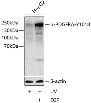
Western blot analysis of lysates from HepG2 cells, using Phospho-PDGFR alpha-Y1018 Rabbit pAb (CABP0615) at 1:1000 dilution. HepG2 cells were treated with UV for 15-30 minutes or treated with EGF (100ng/mL) for 30 minutes after serum-starvation overnight. Secondary antibody: HRP-conjugated Goat anti-Rabbit IgG (H+L) (CABS014) at 1:10000 dilution. Lysates/proteins: 25μg per lane. Blocking buffer: 3% BSA.