The Phospho-PDPK1-S241 Antibody (CABP0426) is a high-quality antibody developed for reliable detection and analysis of target proteins. This antibody, produced in rabbits, is highly specific for the phosphorylated form of PDPK1 at serine 241 and has been validated for use in Western blot applications.PDPK1, also known as 3-phosphoinositide-dependent protein kinase-1, is a key regulator of the PI3K/Akt signaling pathway, which is involved in cell proliferation and survival. Phosphorylation of PDPK1 at serine 241 is known to enhance its kinase activity and its ability to phosphorylate downstream targets, thereby regulating multiple cellular functions.
This antibody is validated for use in WB, IF/ICC, ELISA applications and has demonstrated reactivity against Human, Mouse, Rat samples.
Product Name:
Phospho-PDPK1-S241 Antibody
SKU:
CABP0426
Size:
20μL, 100μL
Reactivity:
Human, Mouse, Rat
Conjugate:
Unconjugated
Immunogen:
Synthetic peptide. This information is considered to be commercially sensitive.
Tested Applications:
WBIF/ICCELISA
Recommended Dilution:
WB
1:500 - 1:2000
IF/ICC
1:100 - 1:200
ELISA
Recommended starting concentration is 1 μg/mL. Please optimize the concentration based on your specific assay requirements.
Enables 3-phosphoinositide-dependent protein kinase activity; phospholipase activator activity; and phospholipase binding activity. Involved in several processes, including cell surface receptor signaling pathway; regulation of protein kinase activity; and regulation of signal transduction. Acts upstream of or within intracellular signal transduction. Located in cell projection; cytosol; and plasma membrane. Implicated in prostate cancer. Biomarker of lung non-small cell carcinoma.
Purification Method
Affinity purification
Gene ID
5170
RRID
AB_2771410
Buffer Information
Store at -20℃. Avoid freeze / thaw cycles. Buffer: PBS containing 50% glycerol, preserved with proclin300 or sodium azide, pH 7.3.
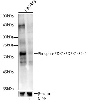
Western blot analysis of lysates from NIH/3T3 cells, using Phospho-PDK1/PDPK1-S241 Rabbit pAb (CABP0426) at 1:1000 dilution. NIH/3T3 cells were treated by λ-PP mixed solution (1ul) at 30℃ for 30 minutes. Secondary antibody: HRP-conjugated Goat anti-Rabbit IgG (H+L) (CABS014) at 1:10000 dilution. Lysates/proteins: 25μg per lane. Blocking buffer: 3% nonfat dry milk in TBST. Detection: ECL Enhanced Kit (AbGn00021). Exposure time: 30s.