The Phospho-PERK-T982 Antibody (CABP0886) is a high-quality antibody developed for reliable detection and analysis of target proteins. This antibody, generated in rabbits, exhibits high specificity for human samples and is optimized for Western blotting applications. It recognizes the phosphorylated form of PERK at threonine 982, enabling precise detection and analysis in various cell types.PERK, also known as PKR-like endoplasmic reticulum kinase, is a crucial component of the unfolded protein response, which helps cells cope with endoplasmic reticulum stress and maintain protein homeostasis.
This antibody is validated for use in WB, IHC-P, ELISA applications and has demonstrated reactivity against Human, Mouse, Rat samples.
Product Name:
Phospho-PERK-T982 Antibody
SKU:
CABP0886
Size:
20μL, 100μL
Reactivity:
Human, Mouse, Rat
Conjugate:
Unconjugated
Immunogen:
Synthetic peptide. This information is considered to be commercially sensitive.
Sequence:
Email for sequence
Tested Applications:
WBIHC-PELISA
Recommended Dilution:
WB
1:500 - 1:1000
IHC-P
1:50 - 1:200
ELISA
Recommended starting concentration is 1 μg/mL. Please optimize the concentration based on your specific assay requirements.
Synonyms:
PEK, WRS, PERK, Phospho-PERK-T982
Positive Sample:
293T treated with IGF-1, A549 treated with FBS
Cellular Localization:
Endoplasmic Reticulum Membrane, Single-Pass Type I Membrane Protein.
Calculated MW:
125kDa
Observed MW:
170kDa
The protein encoded by this gene phosphorylates the alpha subunit of eukaryotic translation-initiation factor 2, leading to its inactivation, and thus to a rapid reduction of translational initiation and repression of global protein synthesis. This protein is thought to modulate mitochondrial function. It is a type I membrane protein located in the endoplasmic reticulum (ER), where it is induced by ER stress caused by malfolded proteins. Mutations in this gene are associated with Wolcott-Rallison syndrome.
Purification Method
Affinity purification
Gene ID
9451
RRID
AB_2771413
Buffer Information
Store at -20℃. Avoid freeze / thaw cycles. Buffer: PBS with 0.09% sodium azide,50% glycerol,pH7.3.
Western blot analysis of lysates from 293T cells, using Phospho-PERK-T982 Rabbit pAb (CABP0886) at 1:1000 dilution. 293T cells were treated with IGF-1 (50 ng/ml) at 37℃ for 5 minutes after serum-starvation overnight. Secondary antibody: HRP-conjugated Goat anti-Rabbit IgG (H+L) (CABS014) at 1:10000 dilution. Lysates/proteins: 25μg per lane. Blocking buffer: 3% BSA. Detection: ECL Basic Kit (AbGn00020). Exposure time: 90s.
Western blot analysis of lysates from A549 cells using Phospho-PERK-T982 Rabbit pAb (CABP0886) at 1:1000 dilution incubated overnight at 4℃. A549 cells were treated with 10% FBS at 37℃ for 5 minutes after serum-starvation overnight. Secondary antibody: HRP-conjugated Goat anti-Rabbit IgG (H+L) (CABS014) at 1:10000 dilution. Lysates/proteins: 30 μg per lane. Blocking buffer: 3 % nonfat dry milk in TBST. Detection: ECL Basic Kit (AbGn00020). Exposure time: 180s.
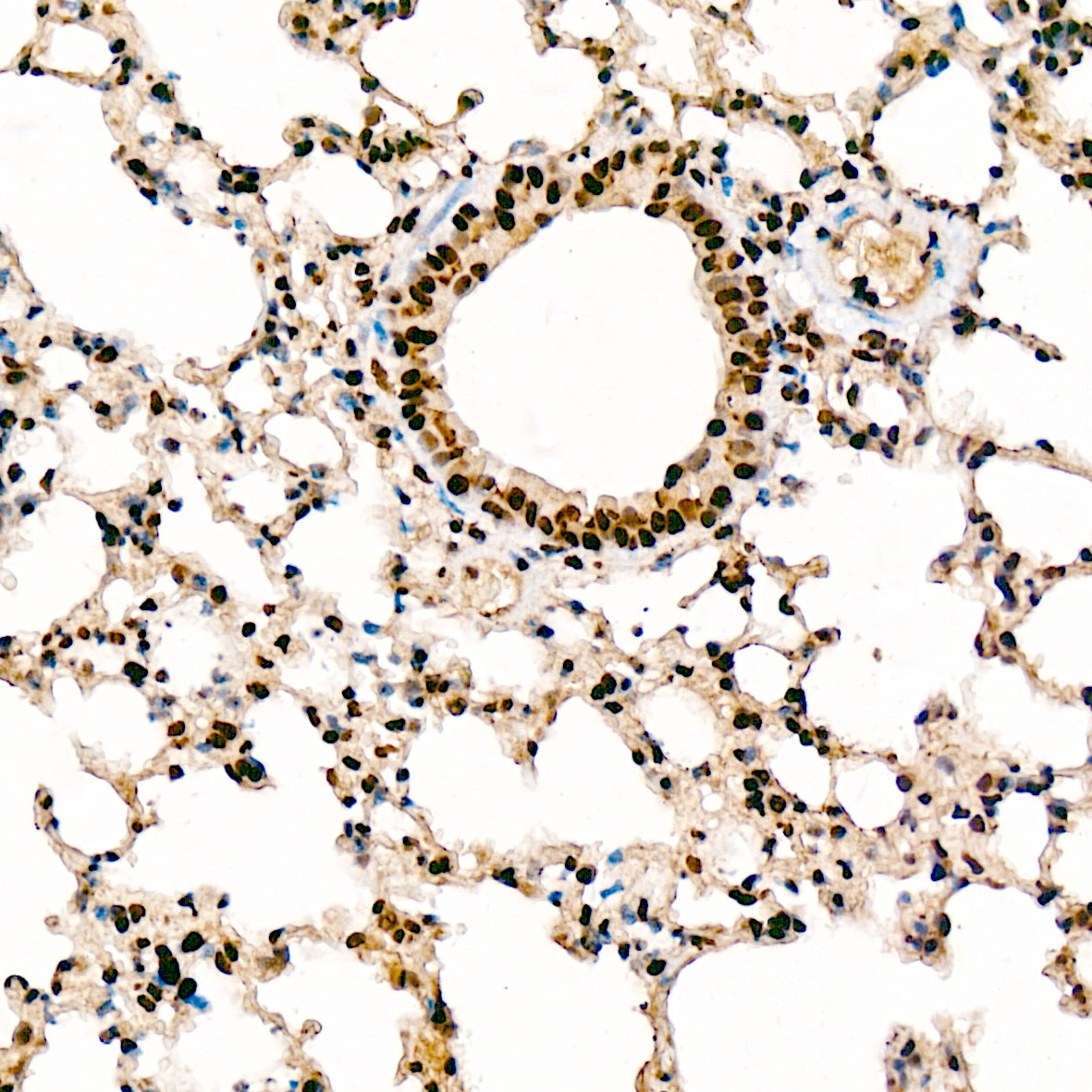
Immunohistochemistry analysis of paraffin-embedded Rat lung tissue using Phospho-PERK-T982 Rabbit pAb (CABP0886) at a dilution of 1:100 (40x lens). High pressure antigen retrieval was performed with 0.01 M citrate buffer (pH 6.0) prior to IHC staining.
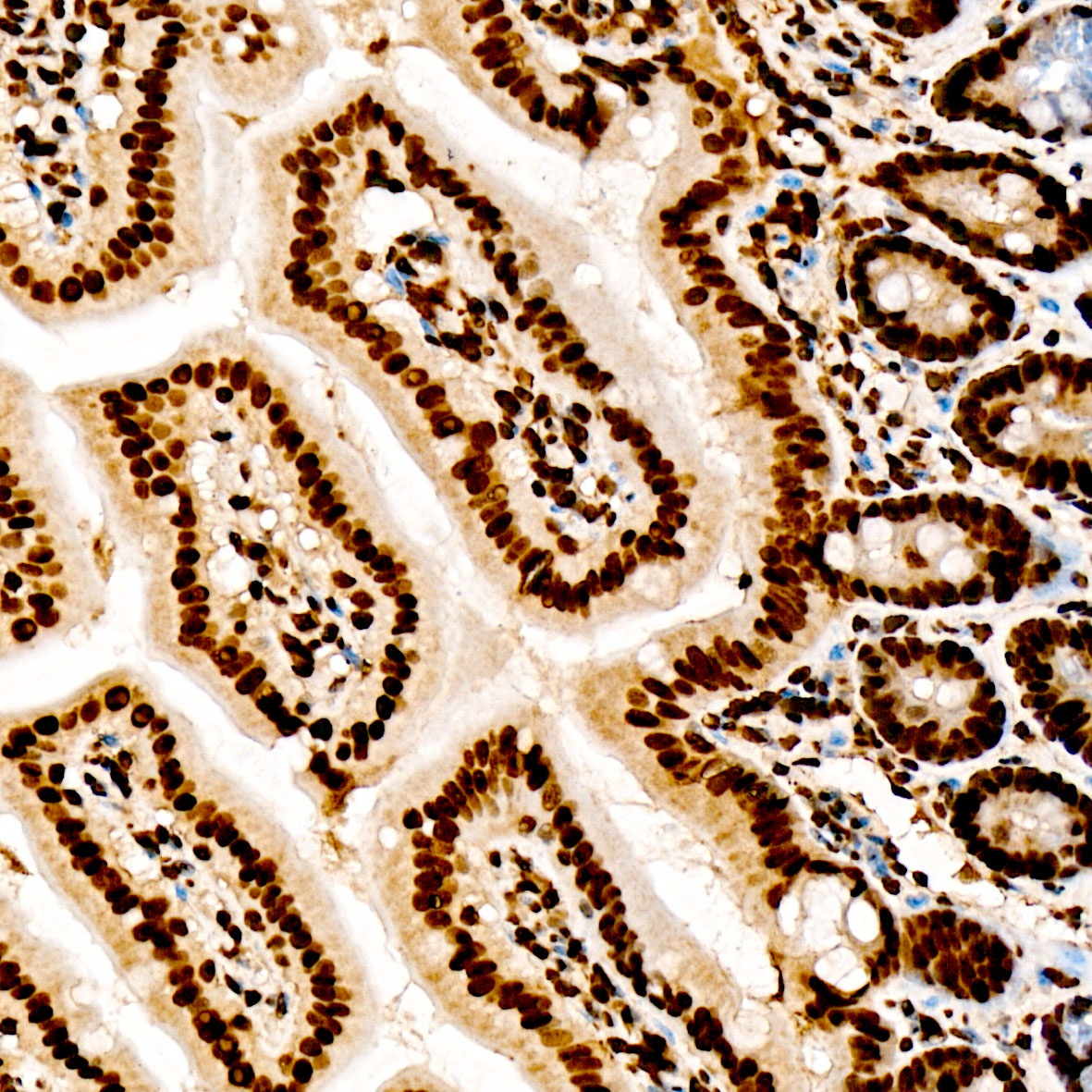
Immunohistochemistry analysis of paraffin-embedded Mouse Intestine tissue using Phospho-PERK-T982 Rabbit pAb (CABP0886) at a dilution of 1:100 (40x lens). High pressure antigen retrieval was performed with 0.01 M citrate buffer (pH 6.0) prior to IHC staining.