The Phospho-NF-kB p65/RelA-T434 Antibody (CABP0838) is a high-quality antibody developed for reliable detection and analysis of target proteins. This antibody, raised in rabbits, exhibits high reactivity with human samples and has been validated for use in Western blot applications. By specifically binding to the phosphorylated form of RelA at threonine 435, this antibody enables precise detection and analysis of this critical post-translational modification in various cell types.The NF-κB signaling pathway plays a crucial role in inflammation, immune response, and cell survival, making it a focal point in research on cancer, autoimmune disorders, and inflammatory diseases.
This antibody is validated for use in WB, ELISA applications and has demonstrated reactivity against Human, Mouse samples.
Product Name:
Phospho-NF-kB p65/RelA-T434 Antibody
SKU:
CABP0838
Size:
20μL, 100μL
Reactivity:
Human, Mouse
Conjugate:
Unconjugated
Immunogen:
Synthetic peptide. This information is considered to be commercially sensitive.
Sequence:
EGTL S
Tested Applications:
WBELISA
Recommended Dilution:
WB
1:500 - 1:2000
ELISA
Recommended starting concentration is 1 μg/mL. Please optimize the concentration based on your specific assay requirements.
NF-kappa-B is a ubiquitous transcription factor involved in several biological processes. It is held in the cytoplasm in an inactive state by specific inhibitors. Upon degradation of the inhibitor, NF-kappa-B moves to the nucleus and activates transcription of specific genes. NF-kappa-B is composed of NFKB1 or NFKB2 bound to either REL, RELA, or RELB. The most abundant form of NF-kappa-B is NFKB1 complexed with the product of this gene, RELA. Four transcript variants encoding different isoforms have been found for this gene.
Purification Method
Affinity purification
Gene ID
5970
RRID
AB_2771514
Buffer Information
Store at -20℃. Avoid freeze / thaw cycles. Buffer: PBS with 0.01% thimerosal,50% glycerol,pH7.3.
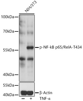
Western blot analysis of lysates from NIH/3T3 cells, using Phospho-NF-kB p65/RelA-T434 pAb (CABP0838) at 1:1000 dilution or RelA antibody (CAB16271). NIH/3T3 cells were treated with TNF-α (20 ng/mL) at 37℃ for 30 minutes. Secondary antibody: HRP-conjugated Goat anti-Rabbit IgG (H+L) (CABS014) at 1:10000 dilution. Lysates/proteins: 25μg per lane. Blocking buffer: 3% BSA. Detection: ECL Basic Kit (AbGn00020). Exposure time: 5min.