The Phospho-Smad2-S465/467 + Smad3-S423/425 Antibody (CABP0548) is a high-quality antibody developed for reliable detection and analysis of target proteins. This antibody, raised in rabbits, is highly specific for phosphorylated Smad2 at serine 465/467 and phosphorylated Smad3 at serine 423/425 in human samples, allowing for accurate detection and analysis in Western blot applications. The TGF-beta signaling pathway is involved in numerous cellular processes, including cell growth, differentiation, and apoptosis. Dysregulation of this pathway has been implicated in various diseases, including cancer, fibrosis, and immune disorders.
This antibody is validated for use in WB, IHC-P, IF/ICC, ELISA applications and has demonstrated reactivity against Human, Mouse, Rat samples.
Product Name:
Phospho-Smad2-S465/467 + Smad3-S423/425 Antibody
SKU:
CABP0548
Size:
20μL, 100μL
Reactivity:
Human, Mouse, Rat
Conjugate:
Unconjugated
Immunogen:
Synthetic peptide. This information is considered to be commercially sensitive.
Sequence:
SSMS
Tested Applications:
WBIHC-PIF/ICCELISA
Recommended Dilution:
WB
1:500 - 1:5000
IF/ICC
1:50 - 1:200
IHC-P
1:50 - 1:200
ELISA
Recommended starting concentration is 1 μg/mL. Please optimize the concentration based on your specific assay requirements.
The protein encoded by this gene belongs to the SMAD, a family of proteins similar to the gene products of the Drosophila gene 'mothers against decapentaplegic' (Mad) and the C. elegans gene Sma. SMAD proteins are signal transducers and transcriptional modulators that mediate multiple signaling pathways. This protein mediates the signal of the transforming growth factor (TGF)-beta, and thus regulates multiple cellular processes, such as cell proliferation, apoptosis, and differentiation. This protein is recruited to the TGF-beta receptors through its interaction with the SMAD anchor for receptor activation (SARA) protein. In response to TGF-beta signal, this protein is phosphorylated by the TGF-beta receptors. The phosphorylation induces the dissociation of this protein with SARA and the association with the family member SMAD4. The association with SMAD4 is important for the translocation of this protein into the nucleus, where it binds to target promoters and forms a transcription repressor complex with other cofactors. This protein can also be phosphorylated by activin type 1 receptor kinase, and mediates the signal from the activin. Alternatively spliced transcript variants have been observed for this gene. [provided by RefSeq, May 2012]
Purification Method
Affinity purification
Gene ID
4087 4088
RRID
AB_2771541
Buffer Information
Store at -20℃. Avoid freeze / thaw cycles. Buffer: PBS with 0.09% sodium azide,50% glycerol,pH7.3.
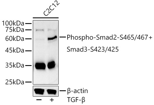
Western blot analysis of lysates from C2C12 cells, using Phospho-Smad2-S465/467 + Smad3-S423/425 Rabbit pAb (CABP0548) at 1:800 dilution. C2C12 cells were treated with TGF-β (10 ng/ml) at 37℃ for 30 minutes. Secondary antibody: HRP-conjugated Goat anti-Rabbit IgG (H+L) (CABS014) at 1:10000 dilution. Lysates/proteins: 25μg per lane. Blocking buffer: 3% nonfat dry milk in TBST. Detection: ECL Basic Kit (AbGn00020). Exposure time: 30s.
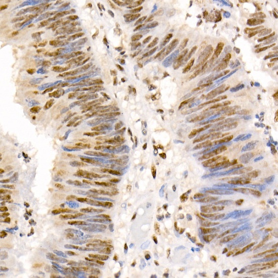
Immunohistochemistry analysis of paraffin-embedded Human colon carcinoma using Phospho-Smad2-S465/467 + Smad3-S423/425 Rabbit pAb (CABP0548) at dilution of 1:50 (40x lens). High pressure antigen retrieval performed with 0.01M Citrate buffer (pH 6.0) prior to IHC staining.
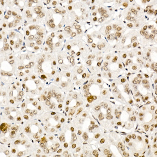
Immunohistochemistry analysis of paraffin-embedded Mouse stomach using Phospho-Smad2-S465/467 + Smad3-S423/425 Rabbit pAb (CABP0548) at dilution of 1:50 (40x lens). High pressure antigen retrieval performed with 0.01M Citrate buffer (pH 6.0) prior to IHC staining.
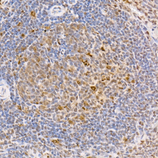
Immunohistochemistry analysis of paraffin-embedded Rat spleen using Phospho-Smad2-S465/467 + Smad3-S423/425 Rabbit pAb (CABP0548) at dilution of 1:50 (40x lens). High pressure antigen retrieval performed with 0.01M Citrate buffer (pH 6.0) prior to IHC staining.
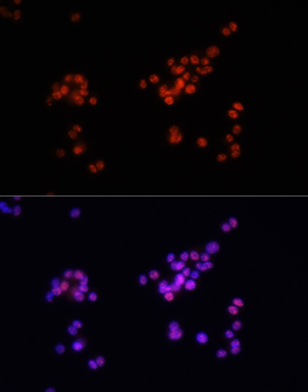
Immunofluorescence analysis of PC-12 cells using Phospho-Smad2-S465/467 + Smad3-S423/425 Rabbit pAb (CABP0548) at dilution of 1:100. Blue: DAPI for nuclear staining.
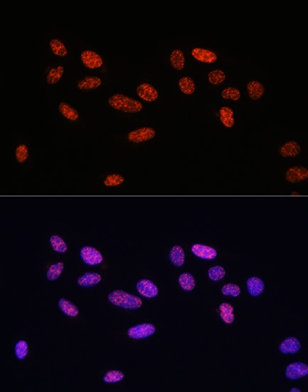
Immunofluorescence analysis of U2OS cells using Phospho-Smad2-S465/467 + Smad3-S423/425 Rabbit pAb (CABP0548) at dilution of 1:100. Blue: DAPI for nuclear staining.