The Phospho-SRC Family-Y416 Antibody (CABP0452) is a high-quality antibody developed for reliable detection and analysis of target proteins. This antibody, raised in rabbits, specifically targets phosphorylated Src at tyrosine 416 and is highly reactive with human samples. Validated for use in Western blot applications, this antibody enables the detection and analysis of activated Src in various cell types.Src is a non-receptor protein tyrosine kinase that plays a crucial role in regulating cell proliferation, survival, and differentiation. Phosphorylation of Src at tyrosine 416 is a critical event that activates its kinase activity and leads to downstream signaling pathways involved in tumorigenesis and metastasis.
This antibody is validated for use in WB, ELISA applications and has demonstrated reactivity against Human, Mouse, Rat samples.
Product Name:
Phospho-SRC Family-Y416 Antibody
SKU:
CABP0452
Size:
20μL, 100μL
Reactivity:
Human, Mouse, Rat
Conjugate:
Unconjugated
Immunogen:
Synthetic peptide. This information is considered to be commercially sensitive.
Sequence:
NEYT A
Tested Applications:
WBELISA
Recommended Dilution:
WB
1:500 - 1:1000
ELISA
Recommended starting concentration is 1 μg/mL. Please optimize the concentration based on your specific assay requirements.
Synonyms:
ASV, SRC1, THC6, c-SRC, p60-Src, SRC, Phospho-SRC Family-Y416
This gene is highly similar to the v-src gene of Rous sarcoma virus. This proto-oncogene may play a role in the regulation of embryonic development and cell growth. The protein encoded by this gene is a tyrosine-protein kinase whose activity can be inhibited by phosphorylation by c-SRC kinase. Mutations in this gene could be involved in the malignant progression of colon cancer. Two transcript variants encoding the same protein have been found for this gene.
Purification Method
Affinity purification
Gene ID
6714 2534 3932 7525 3055 4067
RRID
AB_2771555
Buffer Information
Store at -20℃. Avoid freeze / thaw cycles. Buffer: PBS containing 50% glycerol, preserved with proclin300 or sodium azide, pH 7.3.
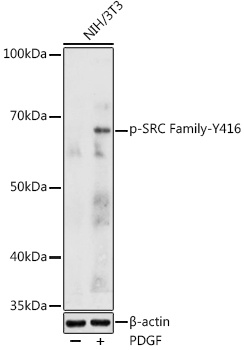
Western blot analysis of lysates from NIH/3T3 cells, using Phospho-SRC Family-Y416 Rabbit pAb (CABP0452) at 1:1000 dilution. NIH/3T3 cells were treated with PDGF (100 ng/ml) at 37℃ for 30 minutes after serum-starvation overnight. Secondary antibody: HRP-conjugated Goat anti-Rabbit IgG (H+L) (CABS014) at 1:10000 dilution. Lysates/proteins: 25μg per lane. Blocking buffer: 3% nonfat dry milk in TBST. Detection: ECL Enhanced Kit (AbGn00021). Exposure time: 180s.