The Phospho-STAT3-S727 Antibody (CABP0474) is a high-quality antibody developed for reliable detection and analysis of target proteins. The protein encoded by this gene is a member of the STAT protein family. In response to cytokines and growth factors, STAT family members are phosphorylated by the receptor associated kinases, and then form homo- or heterodimers that translocate to the cell nucleus where they act as transcription activators. This protein is activated through phosphorylation in response to various cytokines and growth factors including IFNs, EGF, IL5, IL6, HGF, LIF and BMP2. This protein mediates the expression of a variety of genes in response to cell stimuli, and thus plays a key role in many cellular processes such as cell growth and apoptosis. The small GTPase Rac1 has been shown to bind and regulate the activity of this protein. PIAS3 protein is a specific inhibitor of this protein. This gene also plays a role in regulating host response to viral and bacterial infections. Mutations in this gene are associated with infantile-onset multisystem autoimmune disease and hyper-immunoglobulin E syndrome. RRID AB_2771567 Gene ID 6774 Swiss Prot Synonym APRF; HIES; ADMIO; ADMIO1; Phospho-STAT3-S727
This antibody is validated for use in WB, IHC-P, IP, ELISA applications and has demonstrated reactivity against Human, Mouse, Rat samples.
Product Name:
Phospho-STAT3-S727 Antibody
SKU:
CABP0474
Size:
100μL, 20μL
Reactivity:
Human, Mouse, Rat
Clone Number:
-
Conjugate:
Unconjugated
Immunogen:
Synthetic peptide. This information is considered to be commercially sensitive.
Tested Applications:
WBIHC-PIPELISA
Recommended Dilution:
WB
1:500 - 1:5000
IHC-P
1:50 - 1:200
IP
0.5μg-4μg antibody for 200μg-400μg extracts of whole cells
ELISA
Recommended starting concentration is 1 μg/mL. Please optimize the concentration based on your specific assay requirements.
Synonyms:
APRF, HIES, ADMIO, ADMIO1, Phospho-STAT3-S727
Positive Sample:
HeLa, HeLa treated with UV or PMA, HepG2, HepG2 treated with PMA, C6 treated with UV or PMA
Cellular Localization:
Cytoplasm, Nucleus.
Calculated MW:
88kDa
Observed MW:
86kDa
The protein encoded by this gene is a member of the STAT protein family. In response to cytokines and growth factors, STAT family members are phosphorylated by the receptor associated kinases, and then form homo- or heterodimers that translocate to the cell nucleus where they act as transcription activators. This protein is activated through phosphorylation in response to various cytokines and growth factors including IFNs, EGF, IL5, IL6, HGF, LIF and BMP2. This protein mediates the expression of a variety of genes in response to cell stimuli, and thus plays a key role in many cellular processes such as cell growth and apoptosis. The small GTPase Rac1 has been shown to bind and regulate the activity of this protein. PIAS3 protein is a specific inhibitor of this protein. This gene also plays a role in regulating host response to viral and bacterial infections. Mutations in this gene are associated with infantile-onset multisystem autoimmune disease and hyper-immunoglobulin E syndrome. RRID AB_2771567 Gene ID 6774 Swiss Prot Synonym APRF; HIES; ADMIO; ADMIO1; Phospho-STAT3-S727
Purification Method:
Affinity purification
Gene ID:
6774
RRID:
AB_2771567
Buffer Information:
Store at -20℃. Avoid freeze / thaw cycles. Buffer: PBS with 0.09% Sodium azide,50% glycerol,pH7.3.
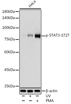
Western blot analysis of lysates from HeLa cells, using Phospho-STAT3-S727 Rabbit pAb (CABP0474) at 1:1000 dilution. HeLa cells were treated with UV at room temperature for 15-30 minutes. HeLa cells were treated with PMA/TPA (200 nM) at 37℃ for 15 minutes after serum-starvation overnight. Secondary antibody: HRP-conjugated Goat anti-Rabbit IgG (H+L) (AS014) at 1:10000 dilution. Lysates/proteins: 25μg per lane. Blocking buffer: 3% nonfat dry milk in TBST. Detection: ECL Basic Kit (AbGn00020). Exposure time: 1s.
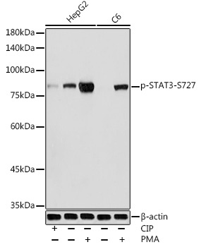
Western blot analysis of various lysates using Phospho-STAT3-S727 Rabbit pAb (CABP0474) at 1:1000 dilution. HepG2 cells and C6 cells were treated with PMA/TPA (200 nM) at 37℃ for 30 minutes after serum-starvation overnight. HepG2 cells were treated with CIP(20uL/400ul) at 37℃ for 1 hour. Secondary antibody: HRP-conjugated Goat anti-Rabbit IgG (H+L) (AS014) at 1:10000 dilution. Lysates/proteins: 25μg per lane. Blocking buffer: 3% nonfat dry milk in TBST. Detection: ECL Basic Kit (AbGn00020). Exposure time: 1s.
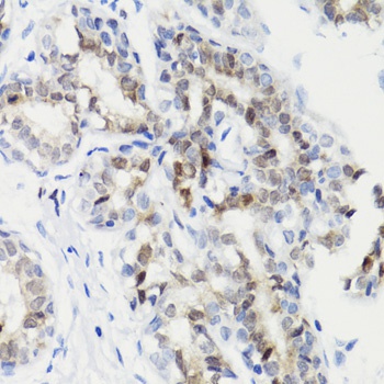
Immunohistochemistry analysis of paraffin-embedded Human breast cancer using Phospho-STAT3-S727 Rabbit pAb (CABP0474) at dilution of 1:100 (40x lens). Microwave antigen retrieval performed with 0.01M Tris/EDTA Buffer (pH 9.0) prior to IHC staining.
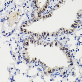
Immunohistochemistry analysis of paraffin-embedded Mouse lung using Phospho-STAT3-S727 Rabbit pAb (CABP0474) at dilution of 1:100 (40x lens). Microwave antigen retrieval performed with 0.01M Tris/EDTA Buffer (pH 9.0) prior to IHC staining.