The Phospho-Stathmin 1-S38 Antibody (CABP0221) is a high-quality antibody developed for reliable detection and analysis of target proteins. Stathmin is a key regulator of microtubule dynamics and plays a critical role in cell proliferation, migration, and cell cycle progression.This antibody, raised in rabbits, is highly specific and has been validated for use in Western blot applications. It recognizes the phosphorylated form of Stathmin at Serine 38, allowing for the detection and quantification of this post-translational modification in cell lysates and tissue samples.Phosphorylation of Stathmin at Serine 38 has been implicated in a variety of cellular processes, including mitotic spindle formation, cytoskeletal organization, and cell motility.
This antibody is validated for use in WB, ELISA applications and has demonstrated reactivity against Human, Mouse, Rat samples.
Product Name:
Phospho-Stathmin 1-S38 Antibody
SKU:
CABP0221
Size:
20μL, 100μL
Reactivity:
Human, Mouse, Rat
Conjugate:
Unconjugated
Immunogen:
Synthetic peptide. This information is considered to be commercially sensitive.
Sequence:
PLSP P
Tested Applications:
WBELISA
Recommended Dilution:
WB
1:500 - 1:2000
ELISA
Recommended starting concentration is 1 μg/mL. Please optimize the concentration based on your specific assay requirements.
This gene belongs to the stathmin family of genes. It encodes a ubiquitous cytosolic phosphoprotein proposed to function as an intracellular relay integrating regulatory signals of the cellular environment. The encoded protein is involved in the regulation of the microtubule filament system by destabilizing microtubules. It prevents assembly and promotes disassembly of microtubules. Multiple transcript variants encoding different isoforms have been found for this gene.
Purification Method
Affinity purification
Gene ID
3925
RRID
AB_2771584
Buffer Information
Store at -20℃. Avoid freeze / thaw cycles. Buffer: PBS containing 50% glycerol, preserved with proclin300 or sodium azide, pH 7.3.
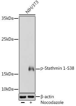
Western blot analysis of lysates from NIH/3T3 cells, using Phospho-Stathmin 1-S38 Rabbit pAb (CABP0221) at 1:1000 dilution. NIH/3T3 cells were treated with Nocodazole (50 ng/ml) at 37℃ for 20 hours. Secondary antibody: HRP-conjugated Goat anti-Rabbit IgG (H+L) (CABS014) at 1:10000 dilution. Lysates/proteins: 25μg per lane. Blocking buffer: 3% BSA. Detection: ECL Basic Kit (AbGn00020). Exposure time: 60s.