The Phospho-Tau-Ser416 Antibody (CABP1101) is a high-quality antibody developed for reliable detection and analysis of target proteins. This antibody, specifically designed for detecting phosphorylation at serine 416 on tau, is highly reactive with human samples and has been validated for use in Western blot applications.Tau protein is commonly associated with neurodegenerative disorders such as Alzheimer's disease, where abnormal phosphorylation leads to the formation of tau tangles in the brain. The Phospho-Tau (Ser416) antibody allows for the detection and analysis of phosphorylated tau in different cell types, providing insights into the role of tau phosphorylation in neurodegeneration.
This antibody is validated for use in WB, IHC-P, ELISA, IF-P applications and has demonstrated reactivity against Human, Mouse, Rat samples.
Product Name:
Phospho-Tau-Ser416 Antibody
SKU:
CABP1101
Size:
20μL, 100μL
Reactivity:
Human, Mouse, Rat
Immunogen:
Synthetic peptide. This information is considered to be commercially sensitive.
Sequence:
TGSI D
Tested Applications:
WBIHC-PELISAIF-P
Recommended Dilution:
WB
1:500 - 1:2000
IF-P
1:50 - 1:200
IHC-P
1:50 - 1:200
ELISA
Recommended starting concentration is 1 μg/mL. Please optimize the concentration based on your specific assay requirements.
This gene encodes the microtubule-associated protein tau (MAPT) whose transcript undergoes complex, regulated alternative splicing, giving rise to several mRNA species. MAPT transcripts are differentially expressed in the nervous system, depending on stage of neuronal maturation and neuron type. MAPT gene mutations have been associated with several neurodegenerative disorders such as Alzheimer's disease, Pick's disease, frontotemporal dementia, cortico-basal degeneration and progressive supranuclear palsy.
Purification Method
Affinity purification
Gene ID
4137
RRID
AB_2863970
Buffer Information
Store at -20℃. Avoid freeze / thaw cycles. Buffer: PBS with 0.01% thimerosal,50% glycerol,pH7.3.
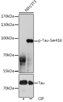
Western blot analysis of lysates from NIH/3T3 cells, using Phospho-Tau-Ser416 Rabbit pAb (CAB0002). NIH/3T3 cells were treated by CIP(20uL/400ul) at 37℃ for 1 hour. Secondary antibody: HRP-conjugated Goat anti-Rabbit IgG (H+L) (CABS014) at 1:10000 dilution. Lysates/proteins: 25μg per lane. Blocking buffer: 3% BSA. Detection: ECL Basic Kit (AbGn00020). Exposure time: 60s.
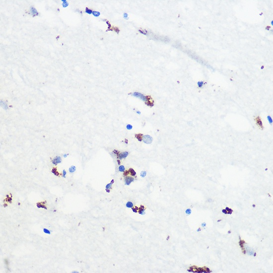
Immunohistochemistry analysis of paraffin-embedded Human brain using Phospho-Tau-Ser416 Rabbit pAb (CABP1101) at dilution of 1:100 (40x lens). Microwave antigen retrieval performed with 0.01M Tris/EDTA Buffer (pH 9.0) prior to IHC staining.
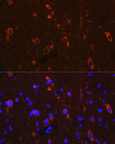
Immunofluorescence analysis of paraffin-embedded Mouse brain using Phospho-Tau-Ser416 Rabbit pAb (CABP1101) at dilution of 1:100. Secondary antibody: Cy3-conjugated Goat anti-Rabbit IgG (H+L) (CABS007) at 1:500 dilution. Blue: DAPI for nuclear staining.