The Phospho-TSC2-T1462 Antibody (CABP0866) is a high-quality antibody developed for reliable detection and analysis of target proteins. This antibody, produced in rabbits, is highly specific to the phosphorylated form of TSC2 at threonine 1462 and has been validated for use in various applications, including Western blotting.TSC2 is a key player in the mTOR signaling pathway, which regulates cell growth and metabolism in response to nutrient availability and growth factors. Phosphorylation of TSC2 at threonine 1462 is known to modulate its function and impact downstream signaling cascades. By targeting this specific phospho-site, researchers can gain insights into the regulatory mechanisms of TSC2 and its role in diseases such as cancer and metabolic disorders.
This antibody is validated for use in WB, ELISA applications and has demonstrated reactivity against Human, Mouse samples.
Product Name:
Phospho-TSC2-T1462 Antibody
SKU:
CABP0866
Size:
20μL, 100μL
Reactivity:
Human, Mouse
Conjugate:
Unconjugated
Immunogen:
Synthetic peptide. This information is considered to be commercially sensitive.
Sequence:
GYTI S
Tested Applications:
WBELISA
Recommended Dilution:
WB
1:500 - 1:1000
ELISA
Recommended starting concentration is 1 μg/mL. Please optimize the concentration based on your specific assay requirements.
Synonyms:
LAM, TSC4, PPP1R160, Phospho-TSC2-T1462
Positive Sample:
MCF7 treated with IGF-1
Cellular Localization:
Cytoplasm, Membrane, Peripheral Membrane Protein.
Calculated MW:
201kDa
Observed MW:
200kDa
This gene is a tumor suppressor gene that encodes the growth inhibitory protein tuberin. Tuberin interacts with hamartin to form the TSC protein complex which functions in the control of cell growth. This TSC protein complex negatively regulates mammalian target of rapamycin complex 1 (mTORC1) signaling which is a major regulator of anabolic cell growth. Mutations in this gene have been associated with tuberous sclerosis and lymphangioleiomyomatosis.
Purification Method
Affinity purification
Gene ID
7249
RRID
AB_2771631
Buffer Information
Store at -20℃. Avoid freeze / thaw cycles. Buffer: PBS with 0.01% thimerosal,50% glycerol,pH7.3.
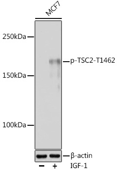
Western blot analysis of lysates from MCF7 cells, using Phospho-TSC2-T1462 Rabbit pAb (CABP0866) at 1:1000 dilution. MCF7 cells were treated with IGF-1 (50 ng/ml) at 37℃ for 5 minutes after serum-starvation overnight. Secondary antibody: HRP-conjugated Goat anti-Rabbit IgG (H+L) (CABS014) at 1:10000 dilution. Lysates/proteins: 25μg per lane. Blocking buffer: 3% nonfat dry milk in TBST. Detection: ECL Basic Kit (AbGn00020). Exposure time: 60s.