The PIK3R3/p55PIK Antibody (CAB17112) is a high-quality antibody developed for reliable detection and analysis of target proteins. This antibody, derived from rabbit origins, exhibits high reactivity with human samples and has been validated for use in Western blot applications.PIK3R3, also known as p55γ, is a critical component in the PI3K signaling pathway, which plays a key role in various cellular processes such as growth, metabolism, and migration. Dysregulation of PI3K signaling has been implicated in numerous diseases, including cancer, diabetes, and cardiovascular disorders.
This antibody is validated for use in WB, IF/ICC, ELISA applications and has demonstrated reactivity against Human, Mouse, Rat samples.
Product Name:
PIK3R3/p55PIK Antibody
SKU:
CAB17112
Size:
20μL, 100μL
Reactivity:
Human, Mouse, Rat
Immunogen:
Synthetic peptide. This information is considered to be commercially sensitive.
Recommended starting concentration is 1 μg/mL. Please optimize the concentration based on your specific assay requirements.
Synonyms:
p55, p55PIK, p55-GAMMA, PIK3R3/p55PIK
Positive Sample:
Mouse testis, Mouse kidney, Rat brain
Cellular Localization:
Cytosol.
Calculated MW:
54kDa
Observed MW:
55kDa
Phosphatidylinositol 3-kinase (PI3K) phosphorylates phosphatidylinositol and similar compounds, which then serve as second messengers in growth signaling pathways. PI3K is composed of a catalytic and a regulatory subunit. The protein encoded by this gene represents a regulatory subunit of PI3K. The encoded protein contains two SH2 domains through which it binds activated protein tyrosine kinases to regulate their activity.
Purification Method
Affinity purification
Gene ID
8503
RRID
AB_2771666
Buffer Information
Store at -20℃. Avoid freeze / thaw cycles. Buffer: PBS with 0.01% thimerosal,50% glycerol,pH7.3.
Western blot analysis of various lysates using PIK3R3/p55PIK Rabbit pAb (CAB17112) at 1:500 dilution. Secondary antibody: HRP-conjugated Goat anti-Rabbit IgG (H+L) (CABS014) at 1:10000 dilution. Lysates/proteins: 25μg per lane. Blocking buffer: 3% nonfat dry milk in TBST. Detection: ECL Basic Kit (AbGn00020). Exposure time: 90s.
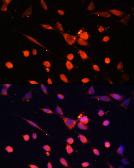
Immunofluorescence analysis of NIH-3T3 cells using PIK3R3/p55PIK Rabbit pAb (CAB17112) at dilution of 1:100. Secondary antibody: Cy3-conjugated Goat anti-Rabbit IgG (H+L) (CABS007) at 1:500 dilution. Blue: DAPI for nuclear staining.

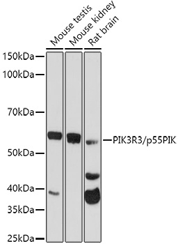


 at 1:10000 dilution. Lysates/proteins: 25ug per lane. Blocking buffer: 3% BSA.")


