The PIK3R4/VPS15 Antibody (CAB15828) is a high-quality antibody developed for reliable detection and analysis of target proteins. This enzyme is involved in regulating various cellular processes, including cell growth, proliferation, and survival. The antibody is produced in rabbits and has high reactivity with human samples, making it suitable for use in Western blot applications.By targeting the PIK3R4 protein, this antibody enables researchers to detect and analyze the expression levels of PI3K in different cell types, providing key insights into its role in cellular signaling pathways.
This antibody is validated for use in WB, IHC-P, IF/ICC, ELISA applications and has demonstrated reactivity against Human, Mouse, Rat samples.
Product Name:
PIK3R4/VPS15 Antibody
SKU:
CAB15828
Size:
20μL, 100μL
Reactivity:
Human, Mouse, Rat
Conjugate:
Unconjugated
Immunogen:
Recombinant protein (or fragment).This information is considered to be commercially sensitive.
Recommended starting concentration is 1 μg/mL. Please optimize the concentration based on your specific assay requirements.
Synonyms:
p150, VPS15, PIK3R4/VPS15
Positive Sample:
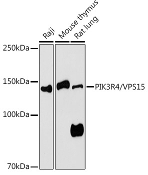
Raji, Mouse thymus, Rat lung
Cellular Localization:
Cytoplasmic Vesicle, Late Endosome, Lipid-Anchor, Membrane, Autophagosome.
Calculated MW:
153kDa
Observed MW:
150kDa
Predicted to enable protein serine/threonine kinase activity. Involved in positive regulation of phosphatidylinositol 3-kinase activity; receptor catabolic process; and regulation of cytokinesis. Located in late endosome and microtubule cytoskeleton.
Purification Method
Affinity purification
Gene ID
30849
RRID
AB_2763252
Buffer Information
Store at -20℃. Avoid freeze / thaw cycles. Buffer: PBS containing 50% glycerol, preserved with proclin300 or sodium azide, pH 7.3.
Western blot analysis of various lysates using PIK3R4/VPS15 Rabbit pAb (CAB15828) at 1:1000 dilution. Secondary antibody: HRP-conjugated Goat anti-Rabbit IgG (H+L) (CABS014) at 1:10000 dilution. Lysates/proteins: 25μg per lane. Blocking buffer: 3% nonfat dry milk in TBST. Detection: ECL Basic Kit (AbGn00020). Exposure time: 180s.
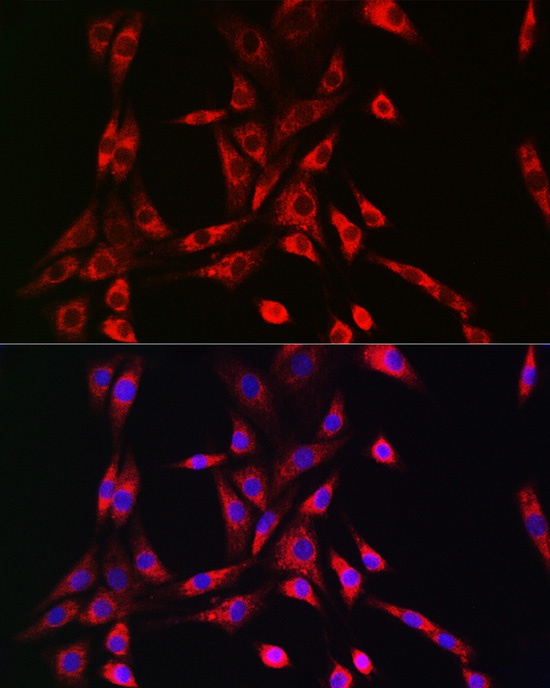
Immunofluorescence analysis of NIH/3T3 cells using PIK3R4/VPS15 Rabbit pAb (CAB15828) at dilution of 1:50 (40x lens). Secondary antibody: Cy3-conjugated Goat anti-Rabbit IgG (H+L) (CABS007) at 1:500 dilution. Blue: DAPI for nuclear staining.