The PKC delta Monoclonal Antibody (CAB7778) is a high-quality antibody developed for reliable detection and analysis of target proteins. This antibody, raised in rabbits, is highly specific for PKC Delta in human samples and is validated for use in Western blot and immunofluorescence applications.PKC Delta is a crucial regulator of cell signaling pathways involved in cancer, cardiovascular diseases, and neurological disorders.
This antibody is validated for use in WB, IHC-P, IF/ICC, ELISA applications and has demonstrated reactivity against Human, Mouse, Rat samples.
Product Name:
PKC delta Monoclonal Antibody
SKU:
CAB7778
Size:
20μL, 100μL
Reactivity:
Human, Mouse, Rat
Clone Number:
ARC1434
Conjugate:
Unconjugated
Immunogen:
Recombinant protein (or fragment).This information is considered to be commercially sensitive.
Sequence:
PFHG DDED ELFE SIRV DTPH YPRW ITKE SKDI LEKL FERE PTKR LGVT GNIK IHPF FKTI NWTL LEKR RLEP PFRP KVKS PRDY SNFD QEFL NEKA RLSY SDKN LID
Tested Applications:
WBIHC-PIF/ICCELISA
Recommended Dilution:
WB
1:500 - 1:2000
IHC-P
1:50 - 1:200
IF/ICC
1:50 - 1:200
ELISA
Recommended starting concentration is 1 μg/mL. Please optimize the concentration based on your specific assay requirements.
The protein encoded by this gene is a member of the protein kinase C family of serine- and threonine-specific protein kinases. The encoded protein is activated by diacylglycerol and is both a tumor suppressor and a positive regulator of cell cycle progression. Also, this protein can positively or negatively regulate apoptosis. Defects in this gene are a cause of autoimmune lymphoproliferative syndrome.
Purification Method
Affinity purification
Gene ID
5580
RRID
AB_2863560
Buffer Information
Store at -20℃. Avoid freeze / thaw cycles. Buffer: PBS containing 50% glycerol and 0.05% BSA, preserved with proclin300 or sodium azide, pH 7.3.
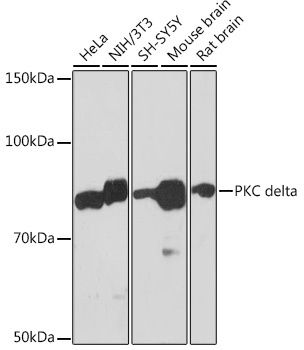
Western blot analysis of various lysates using PKC delta Rabbit mAb (CAB7778) at 1:1000 dilution. Secondary antibody: HRP-conjugated Goat anti-Rabbit IgG (H+L) (CABS014) at 1:10000 dilution. Lysates/proteins: 25μg per lane. Blocking buffer: 3% nonfat dry milk in TBST. Detection: ECL Basic Kit (AbGn00020). Exposure time: 3min.
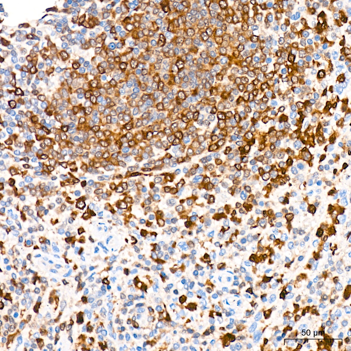
Immunohistochemistry analysis of paraffin-embedded Human spleen tissue using PKC delta Rabbit mAb (CAB7778) at a dilution of 1:200 (40x lens). High pressure antigen retrieval was performed with 0.01 M citrate buffer (pH 6.0) prior to IHC staining.
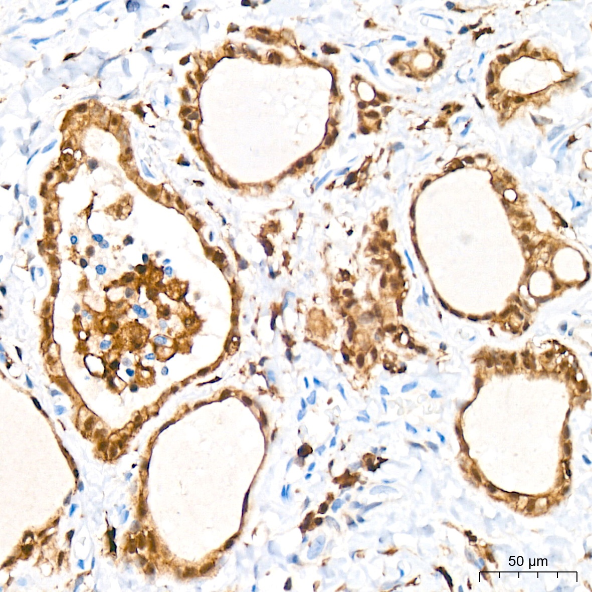
Immunohistochemistry analysis of paraffin-embedded Human thyroid tissue using PKC delta Rabbit mAb (CAB7778) at a dilution of 1:200 (40x lens). High pressure antigen retrieval was performed with 0.01 M citrate buffer (pH 6.0) prior to IHC staining.
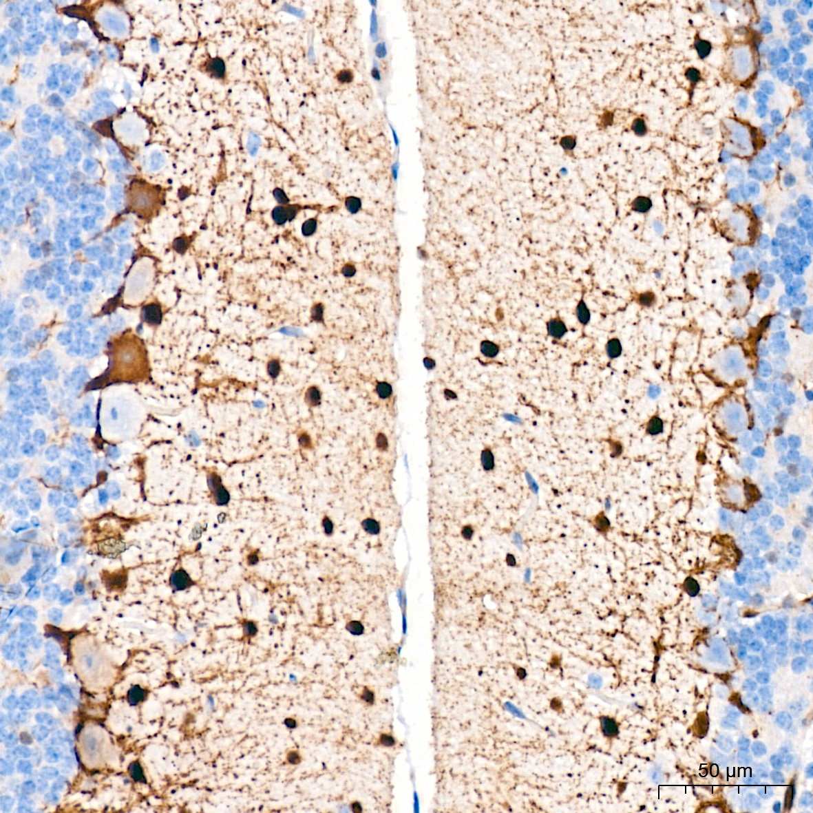
Immunohistochemistry analysis of paraffin-embedded Mouse brain tissue using PKC delta Rabbit mAb (CAB7778) at a dilution of 1:200 (40x lens). High pressure antigen retrieval was performed with 0.01 M citrate buffer (pH 6.0) prior to IHC staining.
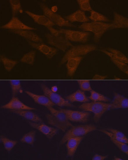
Immunofluorescence analysis of NIH-3T3 cells using PKC delta Rabbit mAb (CAB7778) at dilution of 1:100 (40x lens). Secondary antibody: Cy3-conjugated Goat anti-Rabbit IgG (H+L) (CABS007) at 1:500 dilution. Blue: DAPI for nuclear staining.