The LCP1 Antibody (CAB5561) is a high-quality antibody developed for reliable detection and analysis of target proteins. This antibody, produced in rabbits, has high reactivity with human samples and has been validated for use in Western blot applications. It specifically binds to the Plastin 2 protein, allowing for detection and analysis in a variety of cell types.Plastin 2 plays a crucial role in cell motility and invasion, making it an important target for research in cancer metastasis and other cellular processes.
This antibody is validated for use in WB, IP, ELISA applications and has demonstrated reactivity against Human, Mouse samples.
Product Name:
LCP1 Antibody
SKU:
CAB5561
Size:
20μL, 100μL
Reactivity:
Human, Mouse
Conjugate:
Unconjugated
Immunogen:
Recombinant protein (or fragment).This information is considered to be commercially sensitive.
Plastins are a family of actin-binding proteins that are conserved throughout eukaryote evolution and expressed in most tissues of higher eukaryotes. In humans, two ubiquitous plastin isoforms (L and T) have been identified. Plastin 1 (otherwise known as Fimbrin) is a third distinct plastin isoform which is specifically expressed at high levels in the small intestine. The L isoform is expressed only in hemopoietic cell lineages, while the T isoform has been found in all other normal cells of solid tissues that have replicative potential (fibroblasts, endothelial cells, epithelial cells, melanocytes, etc.). However, L-plastin has been found in many types of malignant human cells of non-hemopoietic origin suggesting that its expression is induced accompanying tumorigenesis in solid tissues.
Purification Method
Affinity purification
Gene ID
3936
RRID
AB_2766341
Buffer Information
Store at -20℃. Avoid freeze / thaw cycles. Buffer: PBS with 0.01% thimerosal,50% glycerol,pH7.3.
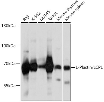
Western blot analysis of various lysates using L-Plastin/LCP1 Rabbit pAb (CAB5561) at 1:1000 dilution. Secondary antibody: HRP-conjugated Goat anti-Rabbit IgG (H+L) (CABS014) at 1:10000 dilution. Lysates/proteins: 25μg per lane. Blocking buffer: 3% nonfat dry milk in TBST. Detection: ECL Basic Kit (AbGn00020). Exposure time: 5s.
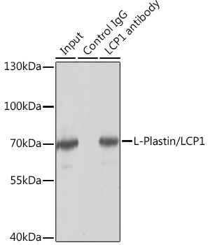
Immunoprecipitation analysis of 150 μg extracts of Jurkat cells using 3 μg L-Plastin/LCP1 antibody (CAB5561). Western blot was performed from the immunoprecipitate using L-Plastin/LCP1 antibody (CAB5561) at a dilution of 1:1000.