The PLP1 Antibody (CAB14251) is a high-quality antibody developed for reliable detection and analysis of target proteins. This antibody, generated in rabbits, exhibits high reactivity with human samples and has been validated for use in Western blot applications.PLP1, also known as proteolipid protein 1, is essential for the formation and maintenance of myelin sheaths, which insulate nerve fibers and facilitate efficient transmission of electrical impulses. Dysregulation of PLP1 is associated with demyelinating diseases such as Pelizaeus-Merzbacher disease and multiple sclerosis, highlighting the importance of studying this protein in neurological research.
This antibody is validated for use in WB, ELISA, IF-P applications and has demonstrated reactivity against Human, Mouse, Rat samples.
Product Name:
PLP1 Antibody
SKU:
CAB14251
Size:
20μL, 100μL
Reactivity:
Human, Mouse, Rat
Conjugate:
Unconjugated
Immunogen:
Recombinant protein (or fragment).This information is considered to be commercially sensitive.
This gene encodes a transmembrane proteolipid protein that is the predominant component of myelin. The encoded protein may play a role in the compaction, stabilization, and maintenance of myelin sheaths, as well as in oligodendrocyte development and axonal survival. Mutations in this gene cause Pelizaeus-Merzbacher disease and spastic paraplegia type 2. Alternatively splicing results in multiple transcript variants, including the DM20 splice variant.
Purification Method
Affinity purification
Gene ID
5354
RRID
AB_2761112
Buffer Information
Store at -20℃. Avoid freeze / thaw cycles. Buffer: PBS containing 50% glycerol, preserved with proclin300 or sodium azide, pH 7.3.
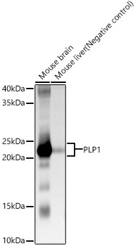
Western blot analysis of various lysates, using PLP1 Rabbit pAb (CAB14251) at 1:1000 dilution. Secondary antibody: HRP-conjugated Goat anti-Rabbit IgG (H+L) (CABS014) at 1:10000 dilution. Lysates/proteins: 25μg per lane. Blocking buffer: 3% nonfat dry milk in TBST. Detection: ECL Basic Kit (AbGn00020). Exposure time: 10s.
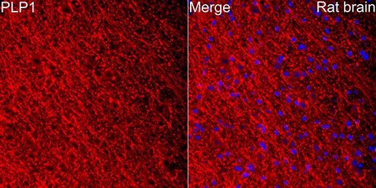
Perform microwave antigen retrieval with 10 mM citrate buffer pH 6.0 before commencing with IF staining protocol.Immunofluorescence analysis of paraffin-embedded rat brain using PLP1 Rabbit pAb (CAB14251) at dilution of 1:100 (40x lens). Secondary antibody: Cy3-conjugated Goat anti-Rabbit IgG (H+L) (CABS007) at 1:500 dilution. Blue: DAPI for nuclear staining.