The PPAT Antibody (CAB6698) is a high-quality antibody developed for reliable detection and analysis of target proteins. This antibody, produced in rabbits, exhibits high reactivity with human samples and has been validated for use in various applications, including Western blot and immunohistochemistry.PPAT, also known as phosphoribosyl pyrophosphate amidotransferase, plays a critical role in the de novo purine biosynthesis pathway, making it a key target for studies in biochemistry and molecular biology. Understanding the function and regulation of PPAT is essential for unraveling the mechanisms underlying purine metabolism and its implications in diseases such as cancer, metabolic disorders, and neurological conditions.
This antibody is validated for use in WB, IF/ICC, ELISA applications and has demonstrated reactivity against Human, Mouse, Rat samples.
Product Name:
PPAT Antibody
SKU:
CAB6698
Size:
20μL, 100μL
Reactivity:
Human, Mouse, Rat
Conjugate:
Unconjugated
Immunogen:
Recombinant protein (or fragment).This information is considered to be commercially sensitive.
Recommended starting concentration is 1 μg/mL. Please optimize the concentration based on your specific assay requirements.
Synonyms:
GPAT, PRAT, ATASE, PPAT
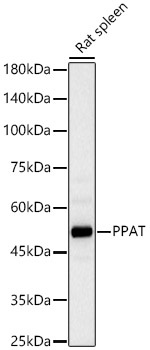
Positive Sample:
Rat spleen
Cellular Localization:
Cytosol.
Calculated MW:
57kDa
Observed MW:
57kDa
The protein encoded by this gene is a member of the purine/pyrimidine phosphoribosyltransferase family. It is a regulatory allosteric enzyme that catalyzes the first step of de novo purine nucleotide biosythetic pathway. This gene and PAICS/AIRC gene, a bifunctional enzyme catalyzing steps six and seven of this pathway, are located in close proximity on chromosome 4, and divergently transcribed from an intergenic region.
Purification Method
Affinity purification
Gene ID
5471
RRID
AB_2767282
Buffer Information
Store at -20℃. Avoid freeze / thaw cycles. Buffer: PBS containing 50% glycerol, preserved with proclin300 or sodium azide, pH 7.3.
Western blot analysis of lysates from Rat spleen, using PPAT Rabbit pAb (CAB6698) at 1:2000 dilution. Secondary antibody: HRP-conjugated Goat anti-Rabbit IgG (H+L) (CABS014) at 1:10000 dilution. Lysates/proteins: 25μg per lane. Blocking buffer: 3% nonfat dry milk in TBST. Detection: ECL Basic Kit (AbGn00020). Exposure time: 60s.
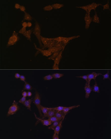
Immunofluorescence analysis of HeLa cells using PPAT Rabbit pAb (CAB6698) at dilution of 1:100 (40x lens). Secondary antibody: Cy3-conjugated Goat anti-Rabbit IgG (H+L) (CABS007) at 1:500 dilution. Blue: DAPI for nuclear staining.