The PRDM14 Antibody (CAB5543) is a high-quality antibody developed for reliable detection and analysis of target proteins. This antibody, produced in rabbits, is highly specific for human samples and has been validated for use in Western blotting applications. By binding to the PRDM14 protein, this antibody enables the detection and analysis of PRDM14 in various cell types, making it an ideal choice for studies in stem cell biology and cancer research.PRDM14 is a transcription factor that plays a crucial role in maintaining the pluripotency of embryonic stem cells and induced pluripotent stem cells.
This antibody is validated for use in WB, IHC-P, ChIP, ELISA applications and has demonstrated reactivity against Human, Mouse, Rat samples.
Product Name:
PRDM14 Antibody
SKU:
CAB5543
Size:
20μL, 100μL
Reactivity:
Human, Mouse, Rat
Conjugate:
Unconjugated
Immunogen:
Recombinant protein (or fragment).This information is considered to be commercially sensitive.
Recommended starting concentration is 1 μg/mL. Please optimize the concentration based on your specific assay requirements.
ChIP
5μg antibody for 10μg-15μg of Chromatin
Synonyms:
PFM11, PRDM14
Positive Sample:
NCI-H125, Mouse ovary, Rat lung
Cellular Localization:
Nucleus.
Calculated MW:
64kDa
Observed MW:
70kDa
This gene encodes a member of the PRDI-BF1 and RIZ homology domain containing (PRDM) family of transcriptional regulators. The encoded protein may possess histone methyltransferase activity and plays a critical role in cell pluripotency by suppressing the expression of differentiation marker genes. Expression of this gene may play a role in breast cancer.
Purification Method
Affinity purification
Gene ID
63978
RRID
AB_2766324
Buffer Information
Store at -20℃. Avoid freeze / thaw cycles. Buffer: PBS containing 50% glycerol, preserved with proclin300 or sodium azide, pH 7.3.
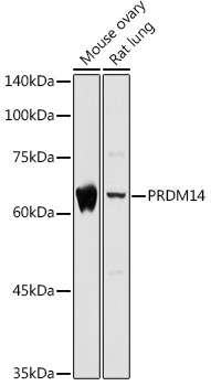
Western blot analysis of various lysates using PRDM14 Rabbit pAb (CAB5543) at 1:1000 dilution. Secondary antibody: HRP-conjugated Goat anti-Rabbit IgG (H+L) (CABS014) at 1:10000 dilution. Lysates/proteins: 25μg per lane. Blocking buffer: 3% nonfat dry milk in TBST. Detection: ECL Basic Kit (AbGn00020). Exposure time: 5s.
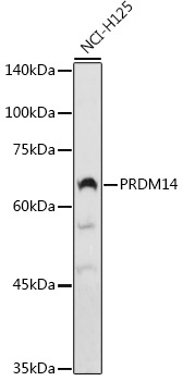
Western blot analysis of lysates from NCI-H125 cells, using PRDM14 Rabbit pAb (CAB5543) at 1:1000 dilution. Secondary antibody: HRP-conjugated Goat anti-Rabbit IgG (H+L) (CABS014) at 1:10000 dilution. Lysates/proteins: 25μg per lane. Blocking buffer: 3% nonfat dry milk in TBST. Detection: ECL Basic Kit (AbGn00020). Exposure time: 90s.
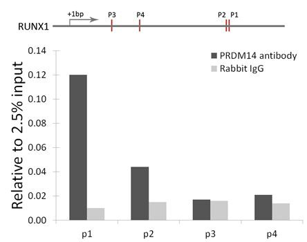
Chromatin immunoprecipitation analysis of RUNX1 gene from 293 cell line, using PRDM14 antibody (CAB5543) and rabbit IgG. P1, P2, P3 and P4 were four probes located on RUNX1 gene as the schematic diagram illustrated. The amount of immunoprecipitated DNA was checked by quantitative PCR. Histogram was constructed by the ratios of the immunoprecipitated DNA to the input.