The AMPKalpha2 Antibody (CAB14052) is a high-quality antibody developed for reliable detection and analysis of target proteins. This antibody is produced in rabbits and is highly specific for human samples, making it ideal for Western blot applications.PRKAA2, also known as AMPKα2, is involved in the regulation of cellular energy homeostasis and metabolism. It plays a crucial role in activating catabolic pathways to generate ATP and in inhibiting anabolic pathways during times of cellular stress.
This antibody is validated for use in WB, IF/ICC, ELISA applications and has demonstrated reactivity against Human, Mouse, Rat samples.
Product Name:
AMPKalpha2 Antibody
SKU:
CAB14052
Size:
20μL, 100μL
Reactivity:
Human, Mouse, Rat
Conjugate:
Unconjugated
Immunogen:
Recombinant protein (or fragment).This information is considered to be commercially sensitive.
Recommended starting concentration is 1 μg/mL. Please optimize the concentration based on your specific assay requirements.
Synonyms:
AMPK, AMPK2, PRKAA, AMPKa2, AMPKα2
Positive Sample:
HeLa, 293T, Mouse brain, Mouse heart, Rat skeletal muscle, Rat liver
Cellular Localization:
Cytoplasm, Nucleus.
Calculated MW:
62kDa
Observed MW:
62kDa
The protein encoded by this gene is a catalytic subunit of the AMP-activated protein kinase (AMPK). AMPK is a heterotrimer consisting of an alpha catalytic subunit, and non-catalytic beta and gamma subunits. AMPK is an important energy-sensing enzyme that monitors cellular energy status. In response to cellular metabolic stresses, AMPK is activated, and thus phosphorylates and inactivates acetyl-CoA carboxylase (ACC) and beta-hydroxy beta-methylglutaryl-CoA reductase (HMGCR), key enzymes involved in regulating de novo biosynthesis of fatty acid and cholesterol. Studies of the mouse counterpart suggest that this catalytic subunit may control whole-body insulin sensitivity and is necessary for maintaining myocardial energy homeostasis during ischemia.
Purification Method
Affinity purification
Gene ID
5563
RRID
AB_2760907
Buffer Information
Store at -20℃. Avoid freeze / thaw cycles. Buffer: PBS containing 50% glycerol, preserved with proclin300 or sodium azide, pH 7.3.
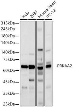
Western blot analysis of various lysates, using AMPKα2 Rabbit pAb (CAB14052) at 1:900 dilution. Secondary antibody: HRP-conjugated Goat anti-Rabbit IgG (H+L) (CABS014) at 1:10000 dilution. Lysates/proteins: 25μg per lane. Blocking buffer: 3% nonfat dry milk in TBST. Detection: ECL Basic Kit (AbGn00020). Exposure time: 10s.
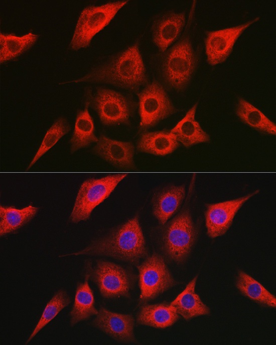
Immunofluorescence analysis of NIH/3T3 cells using AMPKα2 Rabbit pAb (CAB14052) at dilution of 1:100 (40x lens). Secondary antibody: Cy3-conjugated Goat anti-Rabbit IgG (H+L) (CABS007) at 1:500 dilution. Blue: DAPI for nuclear staining.
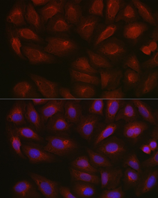
Immunofluorescence analysis of U2OS cells using AMPKα2 Rabbit pAb (CAB14052) at dilution of 1:100 (40x lens). Secondary antibody: Cy3-conjugated Goat anti-Rabbit IgG (H+L) (CABS007) at 1:500 dilution. Blue: DAPI for nuclear staining.
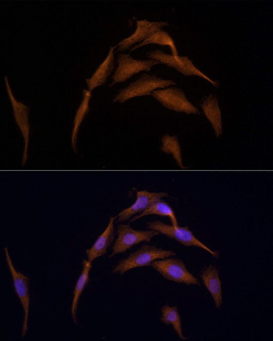
Immunofluorescence analysis of HeLa cells using AMPKα2 Rabbit pAb (CAB14052) at dilution of 1:100 (40x lens). Secondary antibody: Cy3-conjugated Goat anti-Rabbit IgG (H+L) (CABS007) at 1:500 dilution. Blue: DAPI for nuclear staining.