The PRKACB Antibody (CAB5324) is a high-quality antibody developed for reliable detection and analysis of target proteins. The antibody, raised in rabbits, is highly reactive with human samples and has been validated for use in Western blot applications. It binds specifically to the PRKACB protein, allowing for detection and analysis in a variety of cell types, making it an ideal tool for studies in cell signaling and cancer research.PRKACB is involved in the regulation of numerous cellular functions, including metabolism, cell growth, and gene expression. Dysregulation of PRKACB has been implicated in various diseases, making it a potential target for therapeutic intervention.
This antibody is validated for use in WB, IHC-P, IF/ICC, ELISA applications and has demonstrated reactivity against Human, Mouse, Rat samples.
Product Name:
PRKACB Antibody
SKU:
CAB5324
Size:
20μL, 100μL
Reactivity:
Human, Mouse, Rat
Conjugate:
Unconjugated
Immunogen:
Synthetic peptide. This information is considered to be commercially sensitive.
The protein encoded by this gene is a member of the serine/threonine protein kinase family. The encoded protein is a catalytic subunit of cAMP (cyclic AMP)-dependent protein kinase, which mediates signalling though cAMP. cAMP signaling is important to a number of processes, including cell proliferaton and differentiation. Multiple alternatively spliced transcript variants encoding distinct isoforms have been observed.
Purification Method
Affinity purification
Gene ID
5567
RRID
AB_2766136
Buffer Information
Store at -20℃. Avoid freeze / thaw cycles. Buffer: PBS containing 50% glycerol, preserved with proclin300 or sodium azide, pH 7.3.
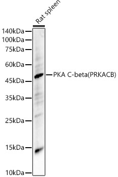
Western blot analysis of lysates from Rat spleen, using PKA C-beta (PRKACB) Rabbit pAb (CAB5324) at 1:1000 dilution. Secondary antibody: HRP-conjugated Goat anti-Rabbit IgG (H+L) (CABS014) at 1:10000 dilution. Lysates/proteins: 25μg per lane. Blocking buffer: 3% nonfat dry milk in TBST. Detection: ECL Basic Kit (AbGn00020). Exposure time: 180s.
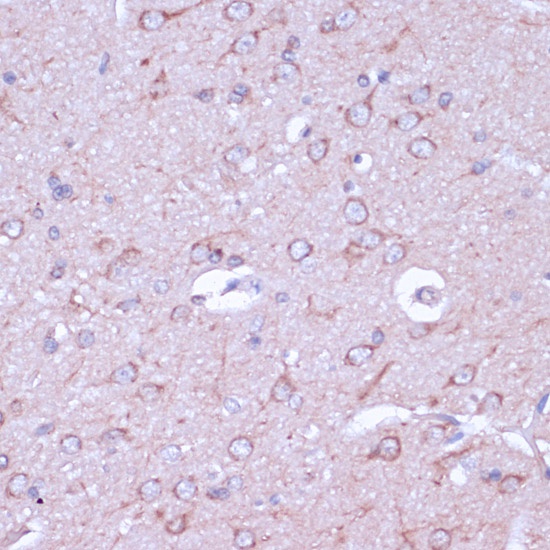
Immunohistochemistry analysis of paraffin-embedded Rat brain using PKA C-beta (PRKACB) Rabbit pAb (CAB5324) at dilution of 1:100 (40x lens). Microwave antigen retrieval performed with 0.01M PBS Buffer (pH 7.2) prior to IHC staining.
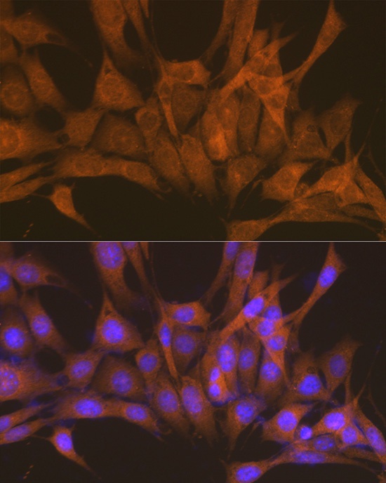
Immunofluorescence analysis of NIH-3T3 cells using PKA C-beta (PRKACB) Rabbit pAb (CAB5324) at dilution of 1:100 (40x lens). Secondary antibody: Cy3-conjugated Goat anti-Rabbit IgG (H+L) (CABS007) at 1:500 dilution. Blue: DAPI for nuclear staining.
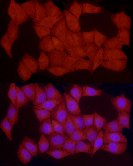
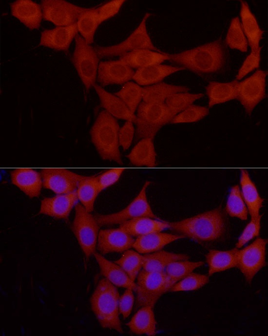
Immunofluorescence analysis of HeLa cells using PKA C-beta (PRKACB) Rabbit pAb (CAB5324) at dilution of 1:50 (40x lens). Secondary antibody: Cy3-conjugated Goat anti-Rabbit IgG (H+L) (CABS007) at 1:500 dilution. Blue: DAPI for nuclear staining.
Immunofluorescence analysis of NIH/3T3 cells using PKA C-beta (PRKACB) Rabbit pAb (CAB5324) at dilution of 1:50 (40x lens). Secondary antibody: Cy3-conjugated Goat anti-Rabbit IgG (H+L) (CABS007) at 1:500 dilution. Blue: DAPI for nuclear staining.