The DR1 Antibody (CAB6903) is a high-quality antibody developed for reliable detection and analysis of target proteins. This antibody, generated in rabbits, exhibits high reactivity with human samples and has been validated for use in Western blot applications. By binding to the DR1 protein, this antibody enables accurate detection and analysis in various cell types, making it ideal for studies in immunology and cancer research.DR1, also known as immune regulatory protein, plays a vital role in maintaining immune balance by modulating inflammatory responses and suppressing immune reactions.
This antibody is validated for use in WB, ELISA applications and has demonstrated reactivity against Human, Mouse, Rat samples.
Product Name:
DR1 Antibody
SKU:
CAB6903
Size:
20μL, 100μL
Reactivity:
Human, Mouse, Rat
Conjugate:
Unconjugated
Immunogen:
Recombinant protein (or fragment).This information is considered to be commercially sensitive.
Recommended starting concentration is 1 μg/mL. Please optimize the concentration based on your specific assay requirements.
Synonyms:
NC2, NC2B, NCB2, NC2-BETA, DR1
Positive Sample:
Mouse testis
Cellular Localization:
Nucleus.
Calculated MW:
19kDa
Observed MW:
19kDa
This gene encodes a TBP- (TATA box-binding protein) associated phosphoprotein that represses both basal and activated levels of transcription. The encoded protein is phosphorylated in vivo and this phosphorylation affects its interaction with TBP. This protein contains a histone fold motif at the amino terminus, a TBP-binding domain, and a glutamine- and alanine-rich region. The binding of DR1 repressor complexes to TBP-promoter complexes may establish a mechanism in which an altered DNA conformation, together with the formation of higher order complexes, inhibits the assembly of the preinitiation complex and controls the rate of RNA polymerase II transcription.
Purification Method
Affinity purification
Gene ID
1810
RRID
AB_2767462
Buffer Information
Store at -20℃. Avoid freeze / thaw cycles. Buffer: PBS containing 50% glycerol, preserved with proclin300 or sodium azide, pH 7.3.
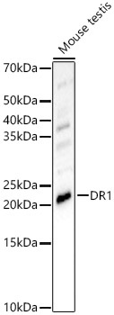
Western blot analysis of lysates from Mouse testis, using DR1 Rabbit pAb (CAB6903) at 1:700 dilution. Secondary antibody: HRP-conjugated Goat anti-Rabbit IgG (H+L) (CABS014) at 1:10000 dilution. Lysates/proteins: 25μg per lane. Blocking buffer: 3% nonfat dry milk in TBST. Detection: ECL Enhanced Kit (AbGn00021). Exposure time: 90s.