The Phospho-MAX-S11 Antibody (CABP0072) is a high-quality antibody developed for reliable detection and analysis of target proteins. This antibody, produced in rabbits, is highly specific to human samples and is validated for use in Western blot applications. By binding to the protein MAX, this antibody enables researchers to detect and analyze protein levels in various cell types, making it an essential tool for studies in cancer biology and cell signaling pathways.
This antibody is validated for use in WB, ELISA applications and has demonstrated reactivity against Human samples.
Product Name:
Phospho-MAX-S11 Antibody
SKU:
CABP0072
Size:
20μL, 100μL
Reactivity:
Human
Conjugate:
Unconjugated
Immunogen:
Synthetic peptide. This information is considered to be commercially sensitive.
Sequence:
VESD E
Tested Applications:
WBELISA
Recommended Dilution:
WB
1:500 - 1:2000
ELISA
Recommended starting concentration is 1 μg/mL. Please optimize the concentration based on your specific assay requirements.
Synonyms:
bHLHd4, Phospho-MAX-S11
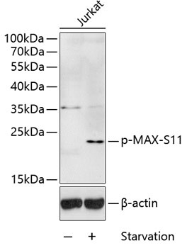
Positive Sample:
Jurkat treated with serum-starvation
Cellular Localization:
Cell Projection, Nucleus, Dendrite.
Calculated MW:
18kDa
Observed MW:
21kDa
The protein encoded by this gene is a member of the basic helix-loop-helix leucine zipper (bHLHZ) family of transcription factors. It is able to form homodimers and heterodimers with other family members, which include Mad, Mxi1 and Myc. Myc is an oncoprotein implicated in cell proliferation, differentiation and apoptosis. The homodimers and heterodimers compete for a common DNA target site (the E box) and rearrangement among these dimer forms provides a complex system of transcriptional regulation. Mutations of this gene have been reported to be associated with hereditary pheochromocytoma. A pseudogene of this gene is located on the long arm of chromosome 7. Alternative splicing results in multiple transcript variants.
Purification Method
Affinity purification
Gene ID
4149
RRID
AB_2771323
Buffer Information
Store at -20℃. Avoid freeze / thaw cycles. Buffer: PBS containing 50% glycerol, preserved with proclin300 or sodium azide, pH 7.3.
Western blot analysis of lysates from Jurkat cells, using Phospho-MAX-S11 Rabbit pAb (CABP0072) at 1:1000 dilution. Jurkat cells were treated with serum-starvation overnight. Secondary antibody: HRP-conjugated Goat anti-Rabbit IgG (H+L) (CABS014) at 1:10000 dilution. Lysates/proteins: 25μg per lane. Blocking buffer: 3% BSA.