The NDRG1 Antibody (CAB2142) is a high-quality antibody developed for reliable detection and analysis of target proteins. This antibody, produced in rabbits, is highly specific and reactive with human samples, making it ideal for use in Western blot applications. By binding to the NDRG1 protein, this antibody allows for the detection and analysis of NDRG1 expression in a variety of cell types.NDRG1, also known as N-myc downstream-regulated gene 1, is a key player in cellular processes such as cell growth, differentiation, and stress response. Its dysregulation has been implicated in various cancers, making it a potential target for cancer therapy and diagnosis.
This antibody is validated for use in WB, IF/ICC, ELISA applications and has demonstrated reactivity against Human, Mouse, Rat samples.
Product Name:
NDRG1 Antibody
SKU:
CAB2142
Size:
20μL, 100μL
Reactivity:
Human, Mouse, Rat
Conjugate:
Unconjugated
Immunogen:
Recombinant protein (or fragment).This information is considered to be commercially sensitive.
This gene is a member of the N-myc downregulated gene family which belongs to the alpha/beta hydrolase superfamily. The protein encoded by this gene is a cytoplasmic protein involved in stress responses, hormone responses, cell growth, and differentiation. The encoded protein is necessary for p53-mediated caspase activation and apoptosis. Mutations in this gene are a cause of Charcot-Marie-Tooth disease type 4D, and expression of this gene may be a prognostic indicator for several types of cancer. Alternatively spliced transcript variants encoding multiple isoforms have been observed for this gene.
Purification Method
Affinity purification
Gene ID
10397
RRID
AB_2764161
Buffer Information
Store at -20℃. Avoid freeze / thaw cycles. Buffer: PBS with 0.09% Sodium azide,50% glycerol,pH7.3.
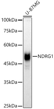
Western blot analysis of lysates from U-87MG cells, using [KO Validated] NDRG1 Rabbit pAb (CAB2142) at 1:1000 dilution. Secondary antibody: HRP-conjugated Goat anti-Rabbit IgG (H+L) (CABS014) at 1:10000 dilution. Lysates/proteins: 25μg per lane. Blocking buffer: 3% nonfat dry milk in TBST. Detection: ECL Basic Kit (AbGn00020). Exposure time: 30s.
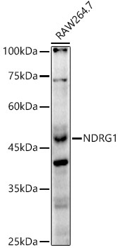
Western blot analysis of lysates from RAW264.7 cells, using [KO Validated] NDRG1 Rabbit pAb (CAB2142) at 1:1000 dilution. Secondary antibody: HRP-conjugated Goat anti-Rabbit IgG (H+L) (CABS014) at 1:10000 dilution. Lysates/proteins: 25μg per lane. Blocking buffer: 3% nonfat dry milk in TBST. Detection: ECL Enhanced Kit (AbGn00021). Exposure time: 120s.
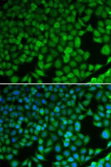
Immunofluorescence analysis of U2OS cells using [KO Validated] NDRG1 Rabbit pAb (CAB2142). Secondary antibody: Cy3-conjugated Goat anti-Rabbit IgG (H+L) (CABS007) at 1:500 dilution. Blue: DAPI for nuclear staining.