The PRPF3 Antibody (CAB5482) is a high-quality antibody developed for reliable detection and analysis of target proteins. This antibody, produced in rabbits, exhibits high reactivity with human samples and has been validated for Western blot applications. By binding specifically to the PRPF3 protein, this antibody enables precise detection and analysis in a variety of cell types, making it an essential reagent for studies in molecular biology and genetic research.PRPF3's key role in the splicing machinery makes it a key player in the regulation of gene expression and the maintenance of cellular homeostasis.
This antibody is validated for use in WB, IHC-P, IF/ICC, IP, ELISA applications and has demonstrated reactivity against Human, Mouse, Rat samples.
Product Name:
PRPF3 Antibody
SKU:
CAB5482
Size:
20μL, 100μL
Reactivity:
Human, Mouse, Rat
Conjugate:
Unconjugated
Immunogen:
Recombinant protein (or fragment).This information is considered to be commercially sensitive.
0.5μg-4μg antibody for 200μg-400μg extracts of whole cells
ELISA
Recommended starting concentration is 1 μg/mL. Please optimize the concentration based on your specific assay requirements.
Synonyms:
PRP3, RP18, HPRP3, Prp3p, HPRP3P, SNRNP90, PRPF3
Positive Sample:
THP-1
Cellular Localization:
Nucleus Speckle.
Calculated MW:
78kDa
Observed MW:
100kDa
The removal of introns from nuclear pre-mRNAs occurs on complexes called spliceosomes, which are made up of 4 small nuclear ribonucleoprotein (snRNP) particles and an undefined number of transiently associated splicing factors. This gene product is one of several proteins that associate with U4 and U6 snRNPs. Mutations in this gene are associated with retinitis pigmentosa-18.
Purification Method
Affinity purification
Gene ID
9129
RRID
AB_2766282
Buffer Information
Store at -20℃. Avoid freeze / thaw cycles. Buffer: PBS containing 50% glycerol, preserved with proclin300 or sodium azide, pH 7.3.
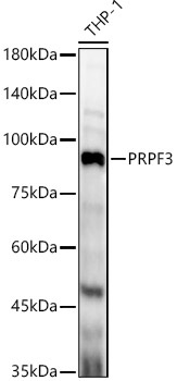
Western blot analysis of lysates from THP-1 cells, using PRPF3 Rabbit pAb (CAB5482) at 1:1000 dilution. Secondary antibody: HRP-conjugated Goat anti-Rabbit IgG (H+L) (CABS014) at 1:10000 dilution. Lysates/proteins: 25μg per lane. Blocking buffer: 3% nonfat dry milk in TBST. Detection: ECL Enhanced Kit (AbGn00021). Exposure time: 60s.
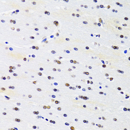
Immunohistochemistry analysis of paraffin-embedded Rat brain using PRPF3 Rabbit pAb (CAB5482) at dilution of 1:100 (40x lens). Microwave antigen retrieval performed with 0.01M PBS Buffer (pH 7.2) prior to IHC staining.
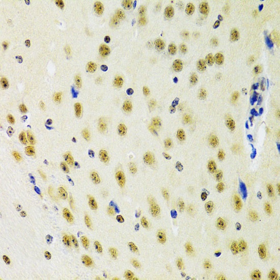
Immunohistochemistry analysis of paraffin-embedded Mouse brain using PRPF3 Rabbit pAb (CAB5482) at dilution of 1:100 (40x lens). Microwave antigen retrieval performed with 0.01M PBS Buffer (pH 7.2) prior to IHC staining.
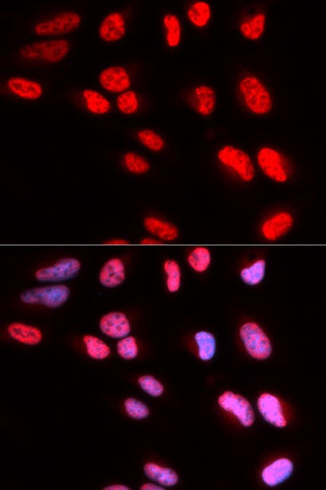
Immunofluorescence analysis of U2OS cells using PRPF3 Rabbit pAb (CAB5482). Secondary antibody: Cy3-conjugated Goat anti-Rabbit IgG (H+L) (CABS007) at 1:500 dilution. Blue: DAPI for nuclear staining.