The PSMA2 Monoclonal Antibody (CAB9182) is a high-quality antibody developed for reliable detection and analysis of target proteins. This antibody, developed using rabbit monoclonal technology, exhibits high reactivity with human samples and has been validated for use in Western blot applications.PSMA2 plays a crucial role in maintaining cellular homeostasis by degrading proteins that are damaged or no longer needed. Dysregulation of the proteasome complex, including PSMA2, has been implicated in various diseases such as cancer, neurodegenerative disorders, and autoimmune conditions.
This antibody is validated for use in WB, IHC-P, IF/ICC, ELISA applications and has demonstrated reactivity against Human, Mouse, Rat samples.
Product Name:
PSMA2 Monoclonal Antibody
SKU:
CAB9182
Size:
20μL, 100μL
Reactivity:
Human, Mouse, Rat
Clone Number:
ARC1465
Conjugate:
Unconjugated
Immunogen:
Recombinant protein (or fragment).This information is considered to be commercially sensitive.
The proteasome is a multicatalytic proteinase complex with a highly ordered ring-shaped 20S core structure. The core structure is composed of 4 rings of 28 non-identical subunits; 2 rings are composed of 7 alpha subunits and 2 rings are composed of 7 beta subunits. Proteasomes are distributed throughout eukaryotic cells at a high concentration and cleave peptides in an ATP/ubiquitin-dependent process in a non-lysosomal pathway. An essential function of a modified proteasome, the immunoproteasome, is the processing of class I MHC peptides. This gene encodes a member of the peptidase T1A family, that is a 20S core alpha subunit.
Purification Method
Affinity purification
Gene ID
5683
RRID
AB_2863680
Buffer Information
Store at -20℃. Avoid freeze / thaw cycles. Buffer: PBS containing 50% glycerol and 0.05% BSA, preserved with proclin300 or sodium azide, pH 7.3.
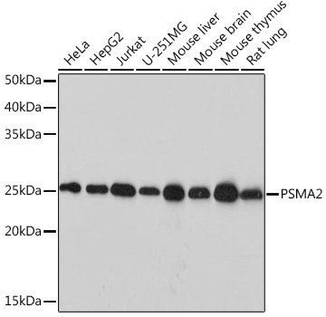
Western blot analysis of various lysates, using PSMA2 Rabbit mAb (CAB9182) at 1:1000 dilution. Secondary antibody: HRP-conjugated Goat anti-Rabbit IgG (H+L) (CABS014) at 1:10000 dilution. Lysates/proteins: 25μg per lane. Blocking buffer: 3% nonfat dry milk in TBST. Detection: ECL Basic Kit (AbGn00020). Exposure time: 10s.
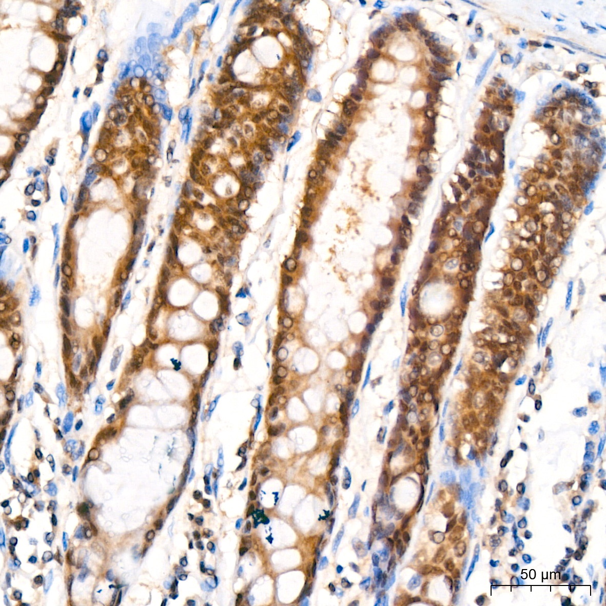
Immunohistochemistry analysis of paraffin-embedded Human colon tissue using PSMA2 Rabbit mAb (CAB9182) at a dilution of 1:200 (40x lens). High pressure antigen retrieval performed with 0.01M Citrate buffer (pH 6.0) prior to IHC staining.
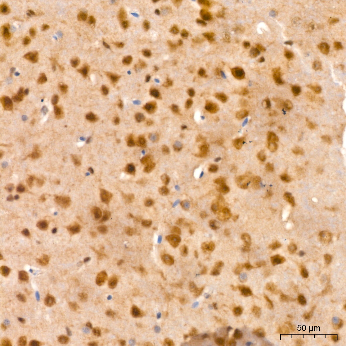
Immunohistochemistry analysis of paraffin-embedded Mouse brain tissue using PSMA2 Rabbit mAb (CAB9182) at a dilution of 1:200 (40x lens). High pressure antigen retrieval performed with 0.01M Citrate buffer (pH 6.0) prior to IHC staining.
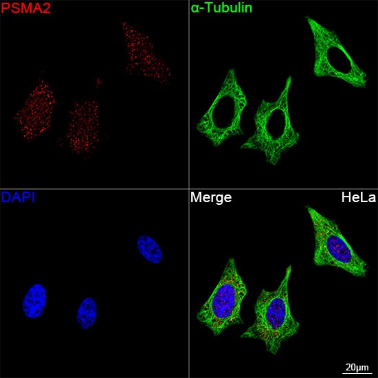
Confocal imaging of HeLa cells using PSMA2 Rabbit mAb (CAB9182, dilution 1:200) followed by a further incubation with Cy3 Goat Anti-Rabbit IgG (H+L) (CABS007, dilution 1:500) (Red). The cells were counterstained with α-Tubulin Mouse mAb (AC012, dilution 1:400) followed by incubation with ABflo® 488-conjugated Goat Anti-Mouse IgG (H+L) Ab (CABS076, dilution 1:500) (Green). DAPI was used for nuclear staining (Blue). Objective: 100x.