The PSMA3 Monoclonal Antibody (CAB5094) is a high-quality antibody developed for reliable detection and analysis of target proteins. This antibody, generated in rabbits, is highly specific for human samples and has been validated for use in Western blot applications. By binding to the PSMA3 protein, this antibody allows for precise detection and analysis in various cell types, making it an excellent choice for studies in molecular biology and cancer research.PSMA3 is a crucial component of the proteasome complex, responsible for degrading unwanted or damaged proteins within cells.
This antibody is validated for use in WB, IHC-P, ELISA applications and has demonstrated reactivity against Human, Mouse, Rat samples.
Product Name:
PSMA3 Monoclonal Antibody
SKU:
CAB5094
Size:
20μL, 100μL
Reactivity:
Human, Mouse, Rat
Clone Number:
ARC1234
Conjugate:
Unconjugated
Immunogen:
Recombinant fusion protein containing a sequence corresponding to amino acids 21-150 of human PSMA3 (P25788).
Recommended starting concentration is 1 μg/mL. Please optimize the concentration based on your specific assay requirements.
Synonyms:
HC8, PSC3, PSMA3
Positive Sample:
HT-29, PC-12, MCF7, NIH/3T3, HL-60, RAW264.7, Rat brain
Cellular Localization:
Cytoplasm, Nucleus.
Calculated MW:
28kDa
Observed MW:
28kDa
The proteasome is a multicatalytic proteinase complex with a highly ordered ring-shaped 20S core structure. The core structure is composed of 4 rings of 28 non-identical subunits; 2 rings are composed of 7 alpha subunits and 2 rings are composed of 7 beta subunits. Proteasomes are distributed throughout eukaryotic cells at a high concentration and cleave peptides in an ATP/ubiquitin-dependent process in a non-lysosomal pathway. An essential function of a modified proteasome, the immunoproteasome, is the processing of class I MHC peptides. This gene encodes a member of the peptidase T1A family, that is a 20S core alpha subunit. Two alternative transcripts encoding different isoforms have been identified.
Purification Method
Affinity purification
Gene ID
5684
RRID
AB_2863441
Buffer Information
Store at -20℃. Avoid freeze / thaw cycles. Buffer: PBS containing 50% glycerol and 0.05% BSA, preserved with proclin300 or sodium azide, pH 7.3.
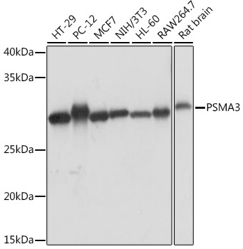
Western blot analysis of various lysates using PSMA3 Rabbit mAb (CAB5094) at 1:1000 dilution. Secondary antibody: HRP-conjugated Goat anti-Rabbit IgG (H+L) (CABS014) at 1:10000 dilution. Lysates/proteins: 25μg per lane. Blocking buffer: 3% nonfat dry milk in TBST. Detection: ECL Basic Kit (AbGn00020). Exposure time: 1s.
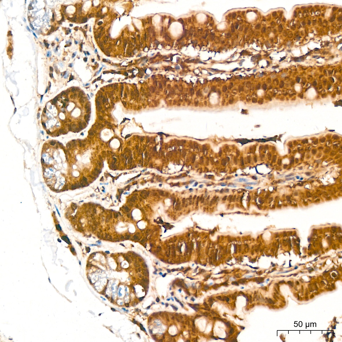
Immunohistochemistry analysis of paraffin-embedded Mouse colon tissue using PSMA3 Rabbit mAb (CAB5094) at a dilution of 1:1000 (40x lens). High pressure antigen retrieval was performed with 0.01 M citrate buffer (pH 6.0) prior to IHC staining.
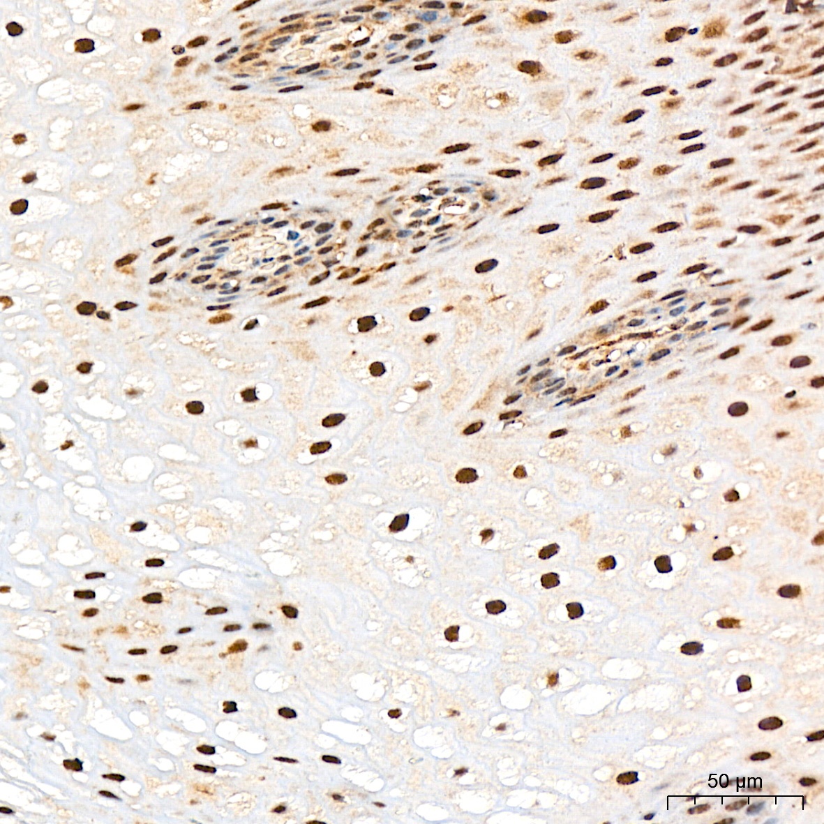
Immunohistochemistry analysis of paraffin-embedded Human esophagus tissue using PSMA3 Rabbit mAb (CAB5094) at a dilution of 1:1000 (40x lens). High pressure antigen retrieval was performed with 0.01 M citrate buffer (pH 6.0) prior to IHC staining.