The PSMD14 Monoclonal Antibody (CAB9608) is a high-quality antibody developed for reliable detection and analysis of target proteins. This antibody, developed using rabbit monoclonal technology, exhibits high specificity and sensitivity towards human samples, making it ideal for use in various biochemical and cell biology assays.PSMD14, also known as Rpn11, is essential for maintaining cellular homeostasis by regulating protein turnover and degradation. Dysregulation of the proteasome system, including PSMD14, has been implicated in various diseases such as cancer, neurodegenerative disorders, and autoimmune conditions.
This antibody is validated for use in WB, IHC-P, IF/ICC, ELISA applications and has demonstrated reactivity against Human, Mouse, Rat samples.
Product Name:
PSMD14 Monoclonal Antibody
SKU:
CAB9608
Size:
20μL, 100μL
Reactivity:
Human, Mouse, Rat
Clone Number:
ARC1655
Conjugate:
Unconjugated
Immunogen:
Recombinant protein (or fragment).This information is considered to be commercially sensitive.
This gene encodes a component of the 26S proteasome. The 26S proteasome is a large multiprotein complex that catalyzes the degradation of ubiquitinated intracellular proteins. The encoded protein is a component of the 19S regulatory cap complex of the 26S proteasome and mediates substrate deubiquitination. A pseudogene of this gene is also located on the long arm of chromosome 2.
Purification Method
Affinity purification
Gene ID
10213
RRID
AB_2863735
Buffer Information
Store at -20℃. Avoid freeze / thaw cycles. Buffer: PBS containing 50% glycerol and 0.05% BSA, preserved with proclin300 or sodium azide, pH 7.3.
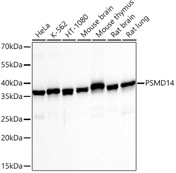
Western blot analysis of various lysates using PSMD14 Rabbit mAb (CAB9608) at 1:1000 dilution incubated overnight at 4℃. Secondary antibody: HRP-conjugated Goat anti-Rabbit IgG (H+L) (CABS014) at 1:10000 dilution. Lysates/proteins: 25 μg per lane. Blocking buffer: 3% nonfat dry milk in TBST. Detection: ECL Basic Kit (AbGn00020). Exposure time: 10s.
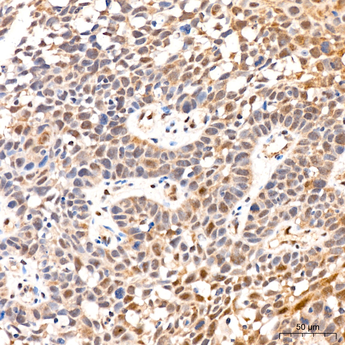
Immunohistochemistry analysis of paraffin-embedded Human cervix cancer tissue using PSMD14 Rabbit mAb (CAB9608) at a dilution of 1:200 (40x lens). High pressure antigen retrieval performed with 0.01M Citrate buffer (pH 6.0) prior to IHC staining.
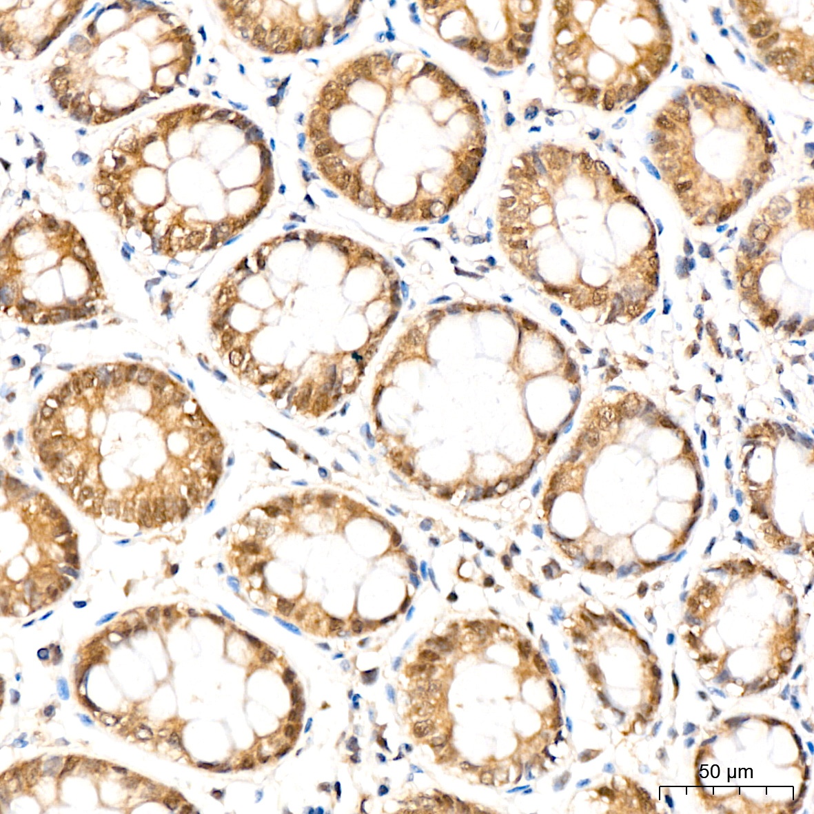
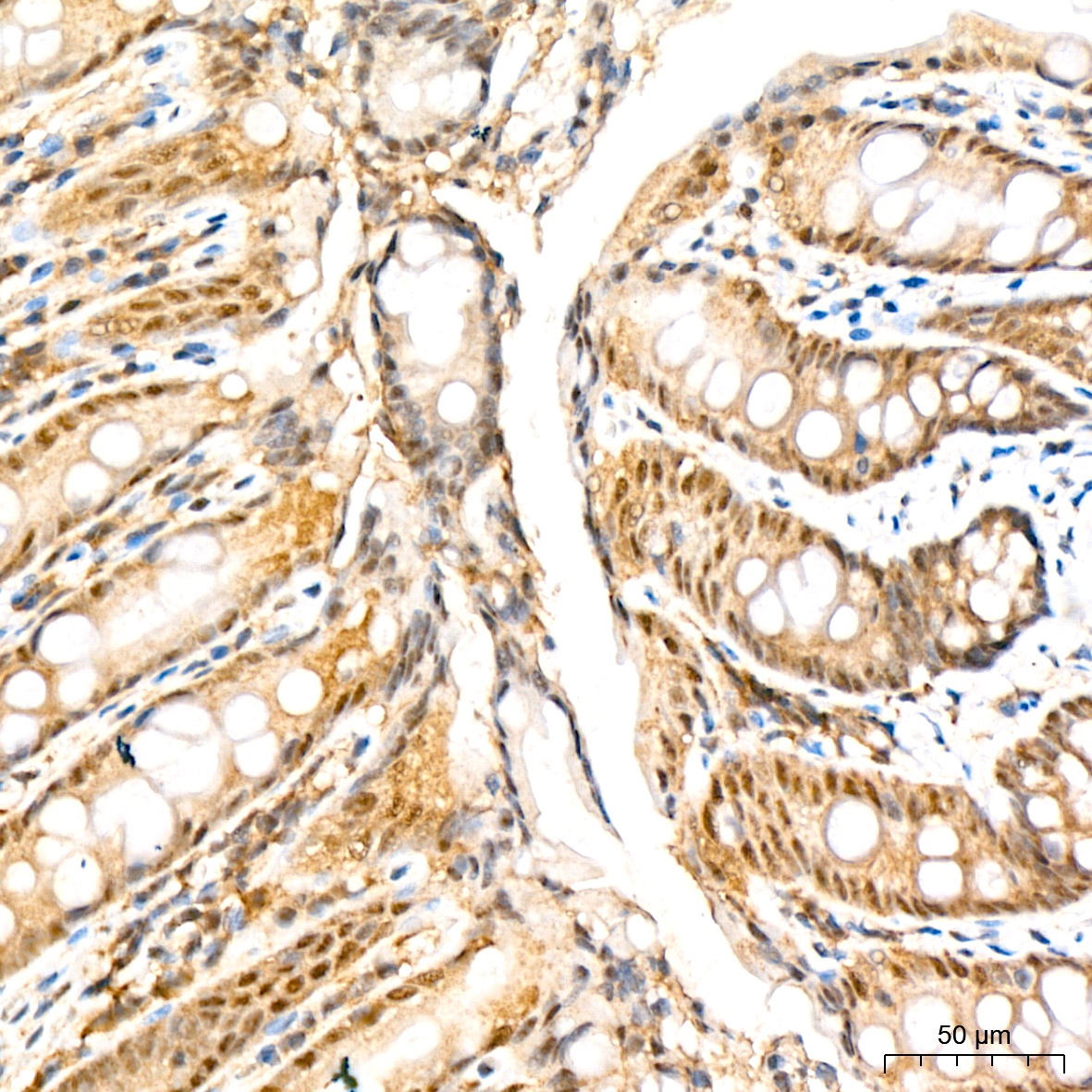
Immunohistochemistry analysis of paraffin-embedded Human colon tissue using PSMD14 Rabbit mAb (CAB9608) at a dilution of 1:200 (40x lens). High pressure antigen retrieval performed with 0.01M Citrate buffer (pH 6.0) prior to IHC staining.
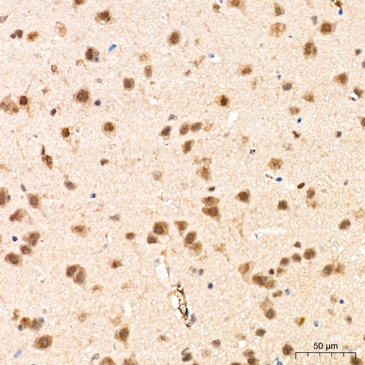
Immunohistochemistry analysis of paraffin-embedded Mouse brain tissue using PSMD14 Rabbit mAb (CAB9608) at a dilution of 1:200 (40x lens). High pressure antigen retrieval performed with 0.01M Citrate buffer (pH 6.0) prior to IHC staining.
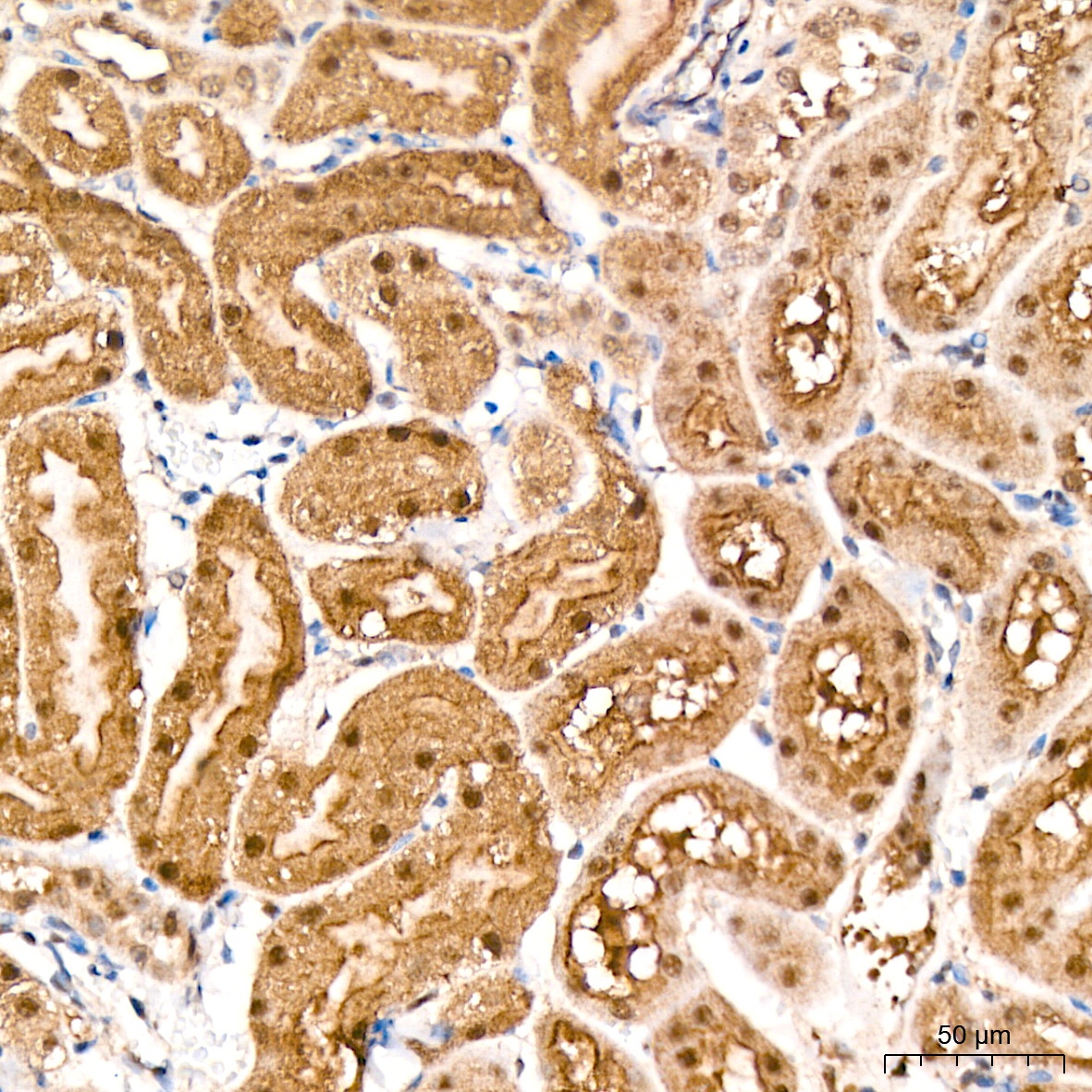
Immunohistochemistry analysis of paraffin-embedded Mouse kidney tissue using PSMD14 Rabbit mAb (CAB9608) at a dilution of 1:200 (40x lens). High pressure antigen retrieval performed with 0.01M Citrate buffer (pH 6.0) prior to IHC staining.
Immunohistochemistry analysis of paraffin-embedded Rat colon tissue using PSMD14 Rabbit mAb (CAB9608) at a dilution of 1:200 (40x lens). High pressure antigen retrieval performed with 0.01M Citrate buffer (pH 6.0) prior to IHC staining.