The FAK Monoclonal Antibody (CAB11131) is a high-quality antibody developed for reliable detection and analysis of target proteins. This antibody, produced through monoclonal antibody technology, specifically targets PTK2 in human samples and is suitable for use in Western blot and immunohistochemistry applications. PTK2, also known as focal adhesion kinase (FAK), plays a crucial role in mediating cell-cell and cell-matrix interactions, making it a key player in cancer metastasis, wound healing, and tissue development.
This antibody is validated for use in WB, IF/ICC, ELISA applications and has demonstrated reactivity against Human, Mouse, Rat samples.
Product Name:
FAK Monoclonal Antibody
SKU:
CAB11131
Size:
20μL, 100μL
Reactivity:
Human, Mouse, Rat
Clone Number:
ARC0171
Conjugate:
Unconjugated
Immunogen:
Synthetic peptide. This information is considered to be commercially sensitive.
This gene encodes a cytoplasmic protein tyrosine kinase which is found concentrated in the focal adhesions that form between cells growing in the presence of extracellular matrix constituents. The encoded protein is a member of the FAK subfamily of protein tyrosine kinases but lacks significant sequence similarity to kinases from other subfamilies. Activation of this gene may be an important early step in cell growth and intracellular signal transduction pathways triggered in response to certain neural peptides or to cell interactions with the extracellular matrix. Several transcript variants encoding different isoforms have been found for this gene.
Purification Method
Affinity purification
Gene ID
5747
RRID
AB_2758423
Buffer Information
Store at -20℃. Avoid freeze / thaw cycles. Buffer: PBS containing 50% glycerol and 0.05% BSA, preserved with proclin300 or sodium azide, pH 7.3.
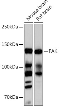
Western blot analysis of various lysates using FAK Rabbit mAb (CAB11131) at 1:1000 dilution. Secondary antibody: HRP-conjugated Goat anti-Rabbit IgG (H+L) (CABS014) at 1:10000 dilution. Lysates/proteins: 25μg per lane. Blocking buffer: 3% nonfat dry milk in TBST. Detection: ECL Basic Kit (AbGn00020). Exposure time: 10s.
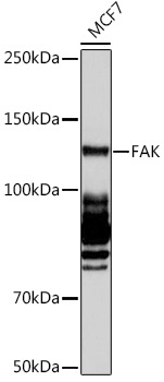
Western blot analysis of lysates from MCF7 cells, using FAK Rabbit mAb (CAB11131) at 1:1000 dilution. Secondary antibody: HRP-conjugated Goat anti-Rabbit IgG (H+L) (CABS014) at 1:10000 dilution. Lysates/proteins: 25μg per lane. Blocking buffer: 3% nonfat dry milk in TBST. Detection: ECL Basic Kit (AbGn00020). Exposure time: 60s.