The ASC/TMS1 Antibody (CAB1170) is a high-quality antibody developed for reliable detection and analysis of target proteins. As a vital component of the inflammasome complex, PYCARD is involved in the activation of caspase-1 and the production of pro-inflammatory cytokines.Produced in rabbits, this antibody is highly specific to human samples and has been validated for use in Western blot applications. By binding to the PYCARD protein, researchers can effectively detect and analyze PYCARD expression in various cell types, making it an ideal tool for studies in immunology, inflammation, and infectious diseases.
This antibody is validated for use in WB, IHC-P, IF/ICC, ELISA, IF-P applications and has demonstrated reactivity against Human, Mouse, Rat samples.
Product Name:
ASC/TMS1 Antibody
SKU:
CAB1170
Size:
20μL, 100μL
Reactivity:
Human, Mouse, Rat
Conjugate:
Unconjugated
Immunogen:
Synthetic peptide. This information is considered to be commercially sensitive.
This gene encodes an adaptor protein that is composed of two protein-protein interaction domains: a N-terminal PYRIN-PAAD-DAPIN domain (PYD) and a C-terminal caspase-recruitment domain (CARD). The PYD and CARD domains are members of the six-helix bundle death domain-fold superfamily that mediates assembly of large signaling complexes in the inflammatory and apoptotic signaling pathways via the activation of caspase. In normal cells, this protein is localized to the cytoplasm; however, in cells undergoing apoptosis, it forms ball-like aggregates near the nuclear periphery. Two transcript variants encoding different isoforms have been found for this gene.
Purification Method
Affinity purification
Gene ID
29108
RRID
AB_2758697
Buffer Information
Store at -20℃. Avoid freeze / thaw cycles. Buffer: PBS containing 50% glycerol, preserved with proclin300 or sodium azide, pH 7.3.
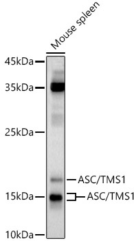
Western blot analysis of lysates from Mouse spleen, using ASC/TMS1 Rabbit pAb (CAB1170) at 1:1000 dilution. Secondary antibody: HRP-conjugated Goat anti-Rabbit IgG (H+L) (CABS014) at 1:10000 dilution. Lysates/proteins: 25μg per lane. Blocking buffer: 3% nonfat dry milk in TBST. Detection: ECL Basic Kit (AbGn00020). Exposure time: 60s.
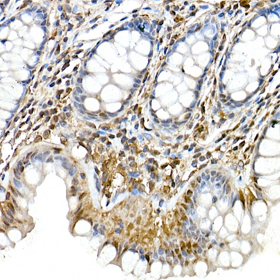
Immunohistochemistry analysis of paraffin-embedded Human colon using ASC/TMS1 Rabbit pAb (CAB1170) at dilution of 1:100 (40x lens). High pressure antigen retrieval performed with 0.01M Citrate buffer (pH 6.0) prior to IHC staining.
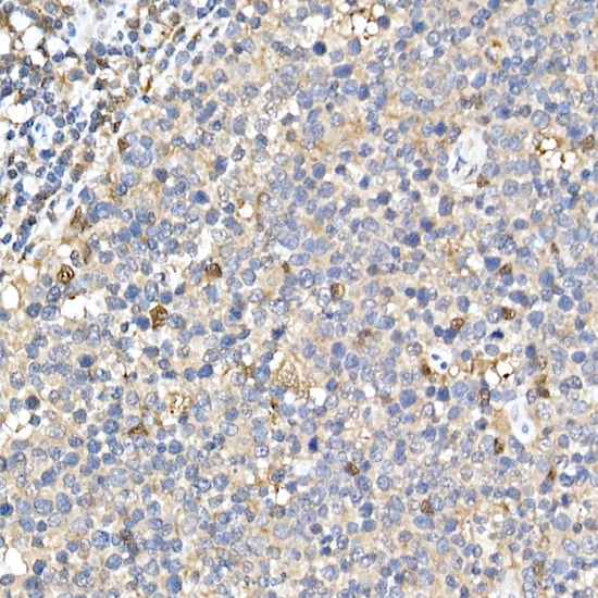
Immunohistochemistry analysis of paraffin-embedded Human tonsil using ASC/TMS1 Rabbit pAb (CAB1170) at dilution of 1:100 (40x lens). High pressure antigen retrieval performed with 0.01M Citrate buffer (pH 6.0) prior to IHC staining.
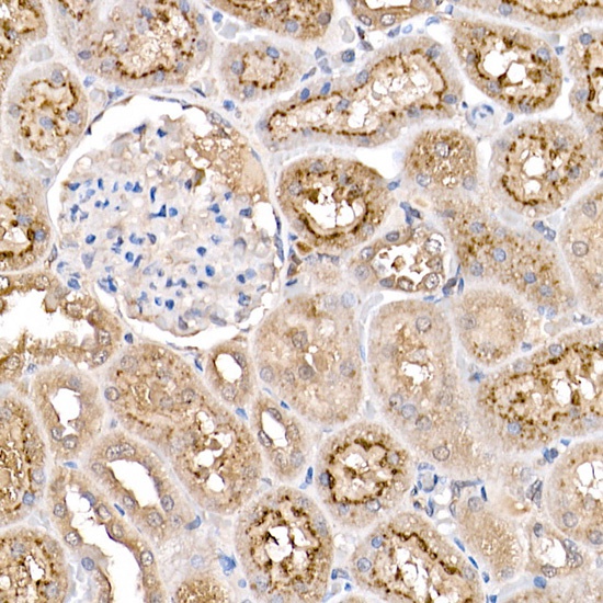
Immunohistochemistry analysis of paraffin-embedded Rat kidney using ASC/TMS1 Rabbit pAb (CAB1170) at dilution of 1:100 (40x lens). High pressure antigen retrieval performed with 0.01M Citrate buffer (pH 6.0) prior to IHC staining.
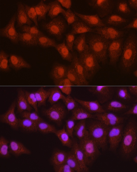
Immunofluorescence analysis of A-549 cells using ASC/TMS1 Rabbit pAb (CAB1170) at dilution of 1:200 (40x lens). Secondary antibody: Cy3-conjugated Goat anti-Rabbit IgG (H+L) (CABS007) at 1:500 dilution. Blue: DAPI for nuclear staining.
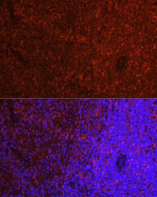
Immunofluorescence analysis of paraffin-embedded rat spleen using ASC/TMS1 Rabbit pAb (CAB1170) at dilution of 1:200 (40x lens). Secondary antibody: Cy3-conjugated Goat anti-Rabbit IgG (H+L) (CABS007) at 1:500 dilution. Blue: DAPI for nuclear staining.