The RAB1A Antibody (CAB14663) is a high-quality antibody developed for reliable detection and analysis of target proteins. This antibody, produced in rabbits, exhibits high reactivity with human samples and is suitable for use in Western blotting applications.Rab1A is a small GTPase that plays a crucial role in maintaining cellular homeostasis by regulating various cellular processes, including protein transport and secretion. Dysregulation of Rab1A has been linked to various diseases, making it a promising target for therapeutic interventions.
This antibody is validated for use in WB, IF/ICC, ELISA applications and has demonstrated reactivity against Mouse, Rat samples.
Product Name:
RAB1A Antibody
SKU:
CAB14663
Size:
20μL, 100μL
Reactivity:
Mouse, Rat
Conjugate:
Unconjugated
Immunogen:
Synthetic peptide. This information is considered to be commercially sensitive.
Recommended starting concentration is 1 μg/mL. Please optimize the concentration based on your specific assay requirements.
Synonyms:
RAB1, YPT1, RAB1A
Positive Sample:
Mouse liver, Mouse lung
Cellular Localization:
Cytoplasm, Early Endosome, Endoplasmic Reticulum, Golgi Apparatus, Melanosome, Membrane, Cytosol.
Calculated MW:
23kDa
Observed MW:
23kDa
This gene encodes a member of the Ras superfamily of GTPases. Members of the gene family cycle between inactive GDP-bound and active GTP-bound forms. This small GTPase controls vesicle traffic from the endoplasmic reticulum to the Golgi apparatus. Multiple alternatively spliced transcript variants have been identified for this gene which encode different protein isoforms.
Purification Method
Affinity purification
Gene ID
5861
RRID
AB_2761539
Buffer Information
Store at -20℃. Avoid freeze / thaw cycles. Buffer: PBS with 0.01% thimerosal,50% glycerol,pH7.3.
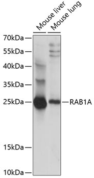
Western blot analysis of various lysates using RAB1A Rabbit pAb (CAB14663) at 1:1000 dilution. Secondary antibody: HRP-conjugated Goat anti-Rabbit IgG (H+L) (CABS014) at 1:10000 dilution. Lysates/proteins: 25μg per lane. Blocking buffer: 3% nonfat dry milk in TBST. Detection: ECL Basic Kit (AbGn00020). Exposure time: 5s.
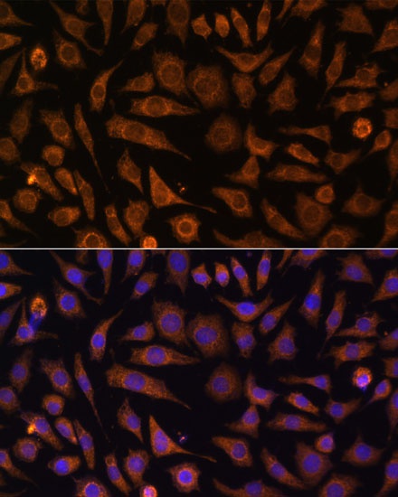
Immunofluorescence analysis of L929 cells using RAB1A Rabbit pAb (CAB14663) at dilution of 1:100. Secondary antibody: Cy3-conjugated Goat anti-Rabbit IgG (H+L) (CABS007) at 1:500 dilution. Blue: DAPI for nuclear staining.