The Raf1 Antibody (CAB0223) is a high-quality antibody developed for reliable detection and analysis of target proteins. This gene is the cellular homolog of viral raf gene (v-raf). The encoded protein is a MAP kinase kinase kinase (MAP3K), which functions downstream of the Ras family of membrane associated GTPases to which it binds directly. Once activated, the cellular RAF1 protein can phosphorylate to activate the dual specificity protein kinases MEK1 and MEK2, which in turn phosphorylate to activate the serine/threonine specific protein kinases, ERK1 and ERK2. Activated ERKs are pleiotropic effectors of cell physiology and play an important role in the control of gene expression involved in the cell division cycle, apoptosis, cell differentiation and cell migration. Mutations in this gene are associated with Noonan syndrome 5 and LEOPARD syndrome 2. RRID AB_2757036 Gene ID 5894 Swiss Prot Synonym NS5; CRAF; Raf-1; c-Raf; CMD1NN; Raf1
This antibody is validated for use in WB, IHC-P, IF/ICC, ELISA applications and has demonstrated reactivity against Human, Mouse, Rat samples.
Product Name:
Raf1 Antibody
SKU:
CAB0223
Size:
100μL, 20μL
Reactivity:
Human, Mouse, Rat
Clone Number:
-
Conjugate:
Unconjugated
Immunogen:
Recombinant protein (or fragment).This information is considered to be commercially sensitive.
Tested Applications:
WBIHC-PIF/ICCELISA
Recommended Dilution:
WB
1:500 - 1:1000
IHC-P
1:50 - 1:200
IF
/
ICC
1:50 - 1:200
ELISA
Recommended starting concentration is 1 μg/mL. Please optimize the concentration based on your specific assay requirements.
Synonyms:
NS5, CRAF, Raf-1, c-Raf, CMD1NN, Raf1
Positive Sample:
Mouse liver
Cellular Localization:
Cell Membrane, Cytoplasm, Mitochondrion, Nucleus.
Calculated MW:
73kDa
Observed MW:
73kDa
This gene is the cellular homolog of viral raf gene (v-raf). The encoded protein is a MAP kinase kinase kinase (MAP3K), which functions downstream of the Ras family of membrane associated GTPases to which it binds directly. Once activated, the cellular RAF1 protein can phosphorylate to activate the dual specificity protein kinases MEK1 and MEK2, which in turn phosphorylate to activate the serine/threonine specific protein kinases, ERK1 and ERK2. Activated ERKs are pleiotropic effectors of cell physiology and play an important role in the control of gene expression involved in the cell division cycle, apoptosis, cell differentiation and cell migration. Mutations in this gene are associated with Noonan syndrome 5 and LEOPARD syndrome 2. RRID AB_2757036 Gene ID 5894 Swiss Prot Synonym NS5; CRAF; Raf-1; c-Raf; CMD1NN; Raf1
Purification Method:
Affinity purification
Gene ID:
5894
RRID:
AB_2757036
Buffer Information:
Store at -20℃. Avoid freeze / thaw cycles. Buffer: PBS containing 50% glycerol, preserved with proclin300 or sodium azide, pH 7.3.
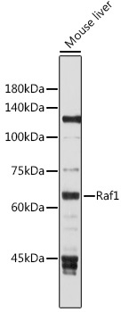
Western blot analysis of lysates from Mouse liver, using Raf1 Rabbit pAb (CAB0223) at 1:1000 dilution. Secondary antibody: HRP-conjugated Goat anti-Rabbit IgG (H+L) (AS014) at 1:10000 dilution. Lysates/proteins: 25μg per lane. Blocking buffer: 3% nonfat dry milk in TBST. Detection: ECL Basic Kit (AbGn00020). Exposure time: 30s.
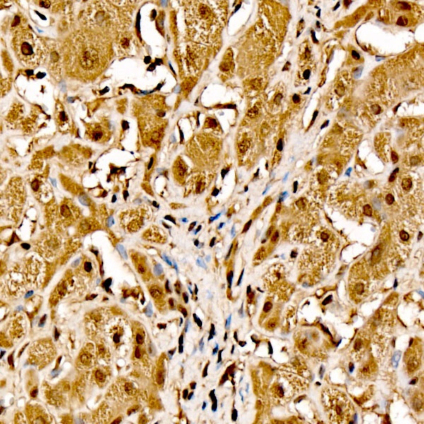
Immunohistochemistry analysis of paraffin-embedded Human liver cancer tissue using Raf1 Rabbit pAb (CAB0223) at a dilution of 1:200 (40x lens). High pressure antigen retrieval performed with 0.01M Citrate buffer (pH 6.0) prior to IHC staining.
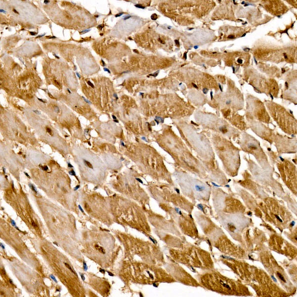
Immunohistochemistry analysis of paraffin-embedded Rat heart tissue using Raf1 Rabbit pAb (CAB0223) at a dilution of 1:200 (40x lens). High pressure antigen retrieval performed with 0.01M Citrate buffer (pH 6.0) prior to IHC staining.
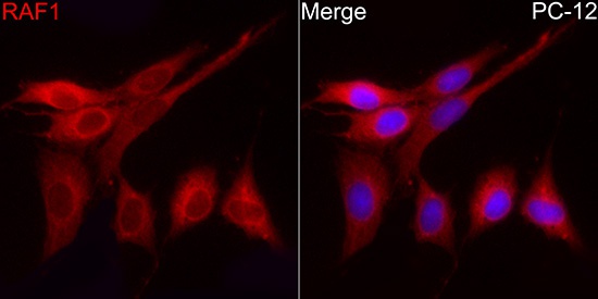
Immunofluorescence analysis of PC-12 cells using Raf1 Rabbit pAb (CAB0223) at dilution of 1:200 (40x lens). Secondary antibody: Cy3-conjugated Goat anti-Rabbit IgG (H+L) (AS007) at 1:500 dilution. Blue: DAPI for nuclear staining.