Description
Rat CYP3A4 (Cytochrome P450 3A4) ELISA Kit
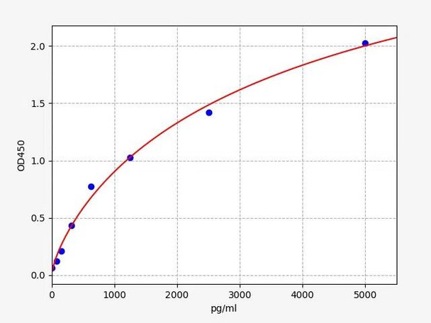
The Rat CYP3A4 (Cytochrome P450 3A4) ELISA Kit (SKU: AEFI03638) is a high-sensitivity, 96T sandwich ELISA assay designed for the quantitative detection of CYP3A4 in rat serum, plasma, cell culture supernatant, tissue and cell lysates, and other biological fluids. Utilizing a double antibody sandwich principle, the assay offers a detection range of 78.125–5000 pg/ml with a sensitivity of 46.875 pg/ml and delivers results within 4 hours. The kit includes pre-coated ELISA microplates, lyophilized standards, biotin-labeled antibodies, HRP-streptavidin conjugate (SABC), TMB substrate, and buffers. Assay precision is excellent, with intra- and inter-assay CVs consistently below 6%. The kit is specific to rat CYP3A4, showing no significant cross-reactivity with related proteins, and is traceable to UniProt ID A0A0P6IZE3.
All reagents are stored at 2–8°C, and the provided manual guides users through sample preparation, reagent dilution, and assay execution. Samples such as serum, plasma, and supernatants are centrifuged and stored as needed, while lysates and homogenates require controlled lysis and clarification steps. The assay procedure involves incubation with standards/samples, biotin-labeled detection antibody, HRP-SABC, and TMB substrate, followed by OD450 measurement. Proper washing (manual or automated) and dilution guidelines help ensure accuracy. A datasheet and MSDS are also available for download, supporting reproducibility and compliance in laboratory environments.
| Product Name: | Rat CYP3A4 (Cytochrome P450 3A4) ELISA Kit |
| SKU: | AEFI03638 |
| Size: | 96T |
| Reactivity: | Rat |
| Range: | 78.125-5000pg/ml |
| Sensitivity: | 46.875pg/ml |
| Detection Method: | Sandwich ELISA, Double Antibody |
| Detection Wavelength: | OD450 |
| Reaction Duration: | 4 hours |
| Synonyms: | CYP3A4 ELISA Kit, Cytochrome P450 3A4 ELISA Kit, CYP3A3 CYPIIIA3 ELISA Kit, CYPIIIA4 ELISA Kit, Nifedipine ELISA Kit, 8-cineole 2-exo-monooxygenase ELISA Kit, Albendazole monooxygenase ELISA Kit, Albendazole sulfoxidase ELISA Kit, Cytochrome P450 3A3 ELISA Kit, Cytochrome P450 HLp ELISA Kit, Cytochrome P450 NF-25 ELISA Kit, Cytochrome P450-PCN1 ELISA Kit |
| Kit Components: |
|
|||||||||||||||||||||||||||||||||||||||||||||||||
Other Materials Required (Not provided)
- Microplate reader (wavelength: 450nm)
- 37°C incubator (CO2 incubator for cell culture is not recommenced.)
- Automated plate washer or multi-channel pipette/5ml pipettor (for manual washing purpose)
- Precision single (0.5-10μL, 5-50μL, 20-200μL, 200-1000μL) and multi-channel pipette with disposable tips(Calibration is required before use.)
- Sterile tubes and Eppendorf tubes with disposable tips
- Absorbent paper and loading slot
- Deionized or distilled water
This assay kit utilizes a double antibody sandwich ELISA method and requires approximately 4 hours to complete. The microplate included in the kit is pre-coated with an anti-CYP3A4 antibody. Standards and appropriately diluted samples are added to the designated wells. Following incubation, unbound substances are removed by washing. A biotinylated detection antibody is then added, which binds specifically to the captured CYP3A4.
After a subsequent wash to remove unbound detection antibody, HRP-conjugated streptavidin (SABC) is added. Following incubation and another wash step, TMB substrate solution is added. The horseradish peroxidase (HRP) catalyzes the conversion of TMB to a blue product, which changes to yellow upon the addition of stop solution.
The optical density (OD) is measured at 450 nm using a microplate reader. The concentration of CYP3A4 in the sample is determined by generating a standard curve, with the OD450 value being directly proportional to the amount of CYP3A4 present.
| Recovery: |
A known quantity of CYP3A4 was spiked into the sample. Recovery was calculated by comparing the measured value with the expected concentration of CYP3A4 in the sample.
|
||||||||||||||||||||||||||||||||||||||||||
| Linearity: |
Samples spiked with CYP3A4 were serially diluted (1:2, 1:4, and 1:8). The linearity was assessed based on the recovery rates at each dilution.
|
||||||||||||||||||||||||||||||||||||||||||
| Precision: |
Intra-assay Precision: Samples with low, medium, and high concentrations were tested 20 times on the same plate. Inter-assay Precision: Samples with low, medium, and high concentrations were tested 20 times across three separate plates.
|
||||||||||||||||||||||||||||||||||||||||||
*Note: Before adding to the wells, equilibrate the TMB substrate for at least 30 mins at room temperature. When diluting samples and reagents, ensure they are mixed completely and evenly. It is recommended to plot a standard curve for each test.
| Step | Protocol |
| 1. | Equilibrate the TMB substrate to room temperature for at least 30 minutes before use. Ensure complete and uniform mixing of all reagents and samples during preparation. |
| 2. | Set up standard, sample, and control (zero) wells on the pre-coated microplate. Record their positions. It is recommended to run each sample and standard in duplicate. |
| 3. | Add 100 µl of standard solution into the designated standard wells. |
| 4. | Add 100 µl of sample dilution buffer into the control (zero) well. |
| 5. | Add 100 µl of properly diluted sample into the test sample wells. |
| 6. | Seal the plate and incubate statically at 37°C for 90 minutes. |
| 7. | Washing: Aspirate the contents and wash the plate 2 times with 350 uL. Do not allow wells to dry. |
| 8. | Add 100 µl of Biotin-labelled Antibody working solution into each well. Avoid touching the well walls. Seal and incubate at 37°C for 60 minutes. |
| 9. | Washing: Wash the plate 3 times with 350 uL. Let the buffer remain in the wells for 1–2 minutes each time. |
| 10. | Add 100 µl of HRP-Streptavidin Conjugate (SABC) working solution into each well. Seal and incubate at 37°C for 30 minutes. |
| 11. | Washing: Wash the plate 5 times with 350 uL. Let the buffer remain in the wells for 1–2 minutes each time. |
| 12. | Add 90 µl of TMB substrate solution into each well. Incubate at 37°C in the dark for 10–20 minutes. Monitor color development; when the highest standard wells turn blue and lower standards show gradual color, proceed to the next step. |
| 13. | Add 50 µl of Stop Solution to each well and mix gently. The color will immediately change to yellow. |
| 14. | Read the optical density (OD) at 450 nm using a microplate reader immediately after adding the stop solution. |
When performing an ELISA assay, correct sample preparation is essential to ensure accurate and reproducible results. Please follow the protocols below based on your sample type.
| Sample Type | Protocol |
| Serum | Place whole blood at room temperature for 2 hours or at 2–8°C overnight. Centrifuge for 20 minutes at 1,000×g and collect the supernatant. Use immediately or aliquot and store at -20°C or -80°C for future assays. |
| Plasma | Use EDTA-Na₂/K₂ as the recommended anticoagulant. Centrifuge for 15 minutes at 1,000×g at 2–8°C within 30 minutes of collection. Collect the supernatant and assay immediately or store at -20°C or -80°C. Note: Over-haemolyzed samples are not suitable for this assay. |
| Cell Culture Supernatant | Collect the culture media, centrifuge at 1,000×g for 20 minutes at 4°C, and collect the clear supernatant. Use immediately or store at -80°C. |
| Cell Lysate |
Suspension Cells:
|
| Tissue Homogenates |
|
| Other Biological Samples | Centrifuge at 1,000×g for 15 minutes at 2–8°C. Use supernatant immediately or store at -80°C for future use. |
| UniProt ID: | A0A0P6IZE3 |
| Sample Type: | Serum, Plasma, Cell Culture Supernatant, Cell or Tissue Lysate, Other Liquid samples |
| Storage: | 2-8°C(Sealed), Don't cryopreserve. |
| Specificity: | Specifically binds with CYP3A4 , no obvious cross reaction with other analogues. |


