The RBL2 Antibody (CAB13649) is a high-quality antibody developed for reliable detection and analysis of target proteins. This antibody, generated in rabbits, exhibits strong reactivity with human samples and has been validated for use in Western blot applications. By binding specifically to the RBL2 protein, researchers can accurately detect and analyze RBL2 expression in a variety of cell types, making it ideal for studies in cancer biology and cell cycle regulation.RBL2, also known as retinoblastoma-like protein 2, plays a crucial role in controlling cell growth and proliferation by inhibiting the activity of cell cycle-promoting factors.
This antibody is validated for use in WB, IF/ICC, ELISA applications and has demonstrated reactivity against Human, Mouse samples.
Product Name:
RBL2 Antibody
SKU:
CAB13649
Size:
20μL, 100μL
Reactivity:
Human, Mouse
Conjugate:
Unconjugated
Immunogen:
Synthetic peptide. This information is considered to be commercially sensitive.
Recommended starting concentration is 1 μg/mL. Please optimize the concentration based on your specific assay requirements.
Synonyms:
Rb2, P130, BRUWAG, RBL2
Positive Sample:
Neuro-2a
Cellular Localization:
Nucleus.
Calculated MW:
128kDa
Observed MW:
130kDa
Enables promoter-specific chromatin binding activity. Involved in regulation of lipid kinase activity. Acts upstream of or within negative regulation of gene expression. Located in chromosome; cytosol; and nuclear lumen.
Purification Method
Affinity purification
Gene ID
5934
RRID
AB_2760511
Buffer Information
Store at -20℃. Avoid freeze / thaw cycles. Buffer: PBS containing 50% glycerol, preserved with proclin300 or sodium azide, pH 7.3.
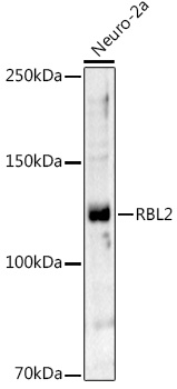
Western blot analysis of lysates from Neuro-2a cells, using RBL2 Rabbit pAb (CAB13649) at 1:1000 dilution. Secondary antibody: HRP-conjugated Goat anti-Rabbit IgG (H+L) (CABS014) at 1:10000 dilution. Lysates/proteins: 25μg per lane. Blocking buffer: 3% nonfat dry milk in TBST. Detection: ECL Basic Kit (AbGn00020). Exposure time: 120s.
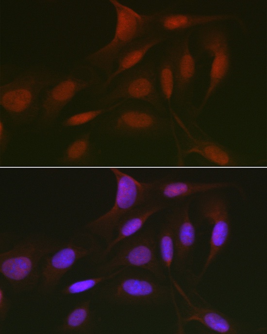
Immunofluorescence analysis of U2OS cells using RBL2 Rabbit pAb (CAB13649) at dilution of 1:50 (40x lens). Secondary antibody: Cy3-conjugated Goat anti-Rabbit IgG (H+L) (CABS007) at 1:500 dilution. Blue: DAPI for nuclear staining.