The RHBDD1 Antibody (CAB14350) is a high-quality antibody developed for reliable detection and analysis of target proteins. Enables serine-type endopeptidase activity. Involved in several processes, including cellular response to unfolded protein; membrane protein proteolysis; and positive regulation of protein catabolic process. Located in endoplasmic reticulum.
This antibody is validated for use in WB, IF/ICC, ELISA applications and has demonstrated reactivity against Human, Mouse, Rat samples.
Product Name:
RHBDD1 Antibody
SKU:
CAB14350
Size:
100μL, 20μL
Reactivity:
Human, Mouse, Rat
Conjugate:
Unconjugated
Immunogen:
Recombinant protein (or fragment).This information is considered to be commercially sensitive.
Tested Applications:
WBIF/ICCELISA
Recommended Dilution:
WB
1:500 - 1:2000
IF/ICC
1:50 - 1:100
ELISA
Recommended starting concentration is 1 μg/mL. Please optimize the concentration based on your specific assay requirements.
Synonyms:
RRP4, RHBDL4, RHBDD1
Positive Sample:
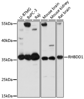
U-87MG, BxPC-3, Raji, mouse brain, mouse kidney, rat brain
Enables serine-type endopeptidase activity. Involved in several processes, including cellular response to unfolded protein; membrane protein proteolysis; and positive regulation of protein catabolic process. Located in endoplasmic reticulum.
Purification Method
Affinity purification
Gene ID
84236
RRID
AB_2761216
Buffer Information
Store at -20℃. Avoid freeze / thaw cycles. Buffer: PBS with 0.01% thimerosal,50% glycerol,pH7.3.
Western blot analysis of various lysates using RHBDD1 Rabbit pAb (CAB14350) at 1:1000 dilution. Secondary antibody: HRP-conjugated Goat anti-Rabbit IgG (H+L) (AS014) at 1:10000 dilution. Lysates/proteins: 25μg per lane. Blocking buffer: 3% nonfat dry milk in TBST. Detection: ECL Basic Kit (AbGn00020). Exposure time: 10s.
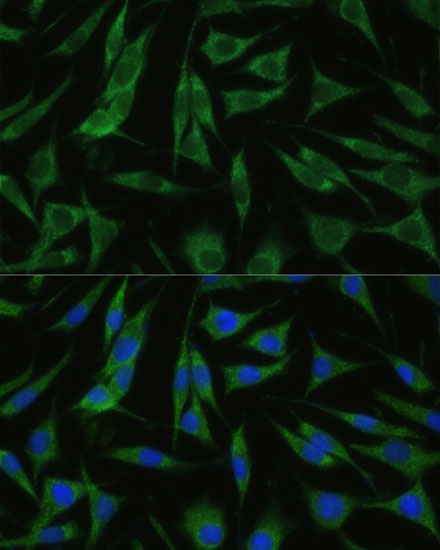
Immunofluorescence analysis of L929 cells using RHBDD1 Rabbit pAb (CAB14350) at dilution of 1:100 (40x lens). Secondary antibody: Cy3-conjugated Goat anti-Rabbit IgG (H+L) (AS007) at 1:500 dilution. Blue: DAPI for nuclear staining.