The RHBDF1 Antibody (CAB17754) is a high-quality antibody developed for reliable detection and analysis of target proteins. This rabbit-derived antibody is highly specific and reacts strongly with human samples, making it an ideal choice for Western blot applications. By binding to RHBDF1, this antibody enables precise detection and analysis of this critical protein in various cellular contexts.RHBDF1, also known as iRhom1, is involved in diverse biological processes such as cell signaling, immune responses, and inflammatory pathways. Dysregulation of RHBDF1 has been implicated in various diseases, including cancer, inflammatory disorders, and neurodegenerative conditions.
This antibody is validated for use in WB, IF/ICC, ELISA applications and has demonstrated reactivity against Mouse, Rat samples.
Product Name:
RHBDF1 Antibody
SKU:
CAB17754
Size:
20μL, 100μL
Reactivity:
Mouse, Rat
Conjugate:
Unconjugated
Immunogen:
Recombinant protein (or fragment).This information is considered to be commercially sensitive.
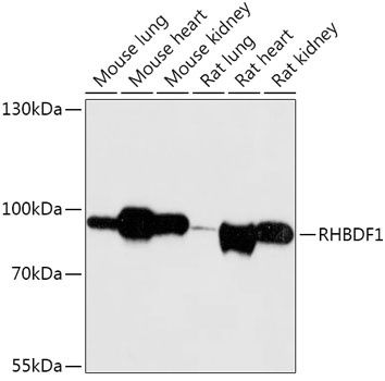
Mouse lung, Mouse heart, Mouse kidney, Rat lung, Rat heart, Rat kidney
Calculated MW:
97kDa
Observed MW:
97kDa
Predicted to enable growth factor binding activity and serine-type endopeptidase activity. Involved in several processes, including negative regulation of protein secretion; regulation of epidermal growth factor receptor signaling pathway; and regulation of proteasomal protein catabolic process. Located in Golgi membrane and endoplasmic reticulum membrane.
Purification Method
Affinity purification
Gene ID
64285
RRID
AB_2772005
Buffer Information
Store at -20℃. Avoid freeze / thaw cycles. Buffer: PBS with 0.01% thimerosal,50% glycerol,pH7.3.
Western blot analysis of various lysates using RHBDF1 Rabbit pAb (CAB17754) at 1:1000 dilution. Secondary antibody: HRP-conjugated Goat anti-Rabbit IgG (H+L) (CABS014) at 1:10000 dilution. Lysates/proteins: 25μg per lane. Blocking buffer: 3% nonfat dry milk in TBST. Detection: ECL Basic Kit (AbGn00020). Exposure time: 30s.
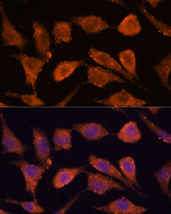
Immunofluorescence analysis of L929 cells using RHBDF1 Rabbit pAb (CAB17754) at dilution of 1:100. Secondary antibody: Cy3-conjugated Goat anti-Rabbit IgG (H+L) (CABS007) at 1:500 dilution. Blue: DAPI for nuclear staining.