The RhoA Antibody (CAB0272) is a high-quality antibody developed for reliable detection and analysis of target proteins. This antibody, raised in rabbits, has high reactivity with human samples and is validated for use in Western blot applications. By specifically binding to RHOA, researchers can effectively detect and analyze the protein in various cell types, making it an essential tool for studies in cell biology and cancer research.RHOA is a member of the Rho family of GTPases and is involved in signaling pathways that regulate cell shape, motility, and proliferation.
This antibody is validated for use in WB, IHC-P, IF/ICC, ELISA applications and has demonstrated reactivity against Human, Mouse, Rat samples.
Product Name:
RhoA Antibody
SKU:
CAB0272
Size:
20μL, 100μL
Reactivity:
Human, Mouse, Rat
Conjugate:
Unconjugated
Immunogen:
Synthetic peptide. This information is considered to be commercially sensitive.
This gene encodes a member of the Rho family of small GTPases, which cycle between inactive GDP-bound and active GTP-bound states and function as molecular switches in signal transduction cascades. Rho proteins promote reorganization of the actin cytoskeleton and regulate cell shape, attachment, and motility. Overexpression of this gene is associated with tumor cell proliferation and metastasis. Multiple alternatively spliced variants have been identified.
Purification Method
Affinity purification
Gene ID
387
RRID
AB_2757085
Buffer Information
Store at -20℃. Avoid freeze / thaw cycles. Buffer: PBS containing 50% glycerol, preserved with proclin300 or sodium azide, pH 7.3.
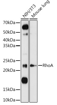
Western blot analysis of various lysates using RhoA Rabbit pAb (CAB0272) at 1:1000 dilution. Secondary antibody: HRP-conjugated Goat anti-Rabbit IgG (H+L) (CABS014) at 1:10000 dilution. Lysates/proteins: 25μg per lane. Blocking buffer: 3% nonfat dry milk in TBST. Detection: ECL Basic Kit (AbGn00020). Exposure time: 90s.
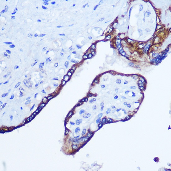
Immunohistochemistry analysis of paraffin-embedded Human placenta using RhoA Rabbit pAb (CAB0272) at dilution of 1:100 (40x lens). Microwave antigen retrieval performed with 0.01M Tris/EDTA Buffer (pH 9.0) prior to IHC staining.
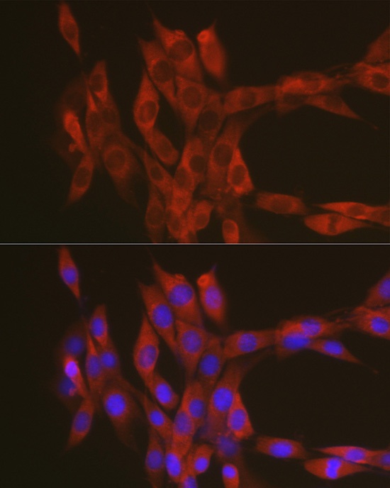
Immunofluorescence analysis of NIH-3T3 cells using RhoA Rabbit pAb (CAB0272) at dilution of 1:100 (40x lens). Secondary antibody: Cy3-conjugated Goat anti-Rabbit IgG (H+L) (CABS007) at 1:500 dilution. Blue: DAPI for nuclear staining.