The RhoB Antibody (CAB2819) is a high-quality antibody developed for reliable detection and analysis of target proteins. This antibody, produced in rabbits, is highly specific for human samples and has been validated for use in Western blotting applications. By binding to RhoB, this antibody allows for the detection and analysis of RhoB expression in various cell types.RhoB plays a crucial role in cellular processes such as cell migration, cell adhesion, and cell cycle regulation. Dysregulation of RhoB has been implicated in cancer progression, making it an attractive target for cancer research.
This antibody is validated for use in WB, ELISA applications and has demonstrated reactivity against Human, Mouse, Rat samples.
Product Name:
RhoB Antibody
SKU:
CAB2819
Size:
20μL, 100μL
Reactivity:
Human, Mouse, Rat
Conjugate:
Unconjugated
Immunogen:
Synthetic peptide. This information is considered to be commercially sensitive.
Recommended starting concentration is 1 μg/mL. Please optimize the concentration based on your specific assay requirements.
Synonyms:
ARH6, ARHB, RHOH6, MST081, MSTP081, RhoB
Positive Sample:
HeLa, HCT116, SK-BR-3, Daudi, Mouse brain, Rat lung
Cellular Localization:
Cell Membrane, Cleavage Furrow, Late Endosome Membrane, Lipid-Anchor, Nucleus.
Calculated MW:
22kDa
Observed MW:
21kDa
Predicted to enable GTP binding activity; GTPase activity; and protein kinase binding activity. Involved in several processes, including cellular response to hydrogen peroxide; cellular response to ionizing radiation; and regulation of cell migration. Located in cleavage furrow and endosome membrane. Biomarker of breast cancer.
Purification Method
Affinity purification
Gene ID
388
RRID
AB_2764656
Buffer Information
Store at -20℃. Avoid freeze / thaw cycles. Buffer: PBS containing 50% glycerol, preserved with proclin300 or sodium azide, pH 7.3.
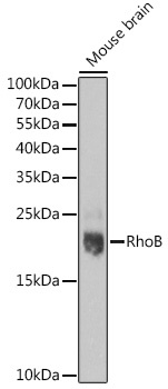
Western blot analysis of lysates from Mouse brain, using RhoB pAb (CAB2819) at 1:1000 dilution. Secondary antibody: HRP-conjugated Goat anti-Rabbit IgG (H+L) (CABS014) at 1:10000 dilution. Lysates/proteins: 25μg per lane. Blocking buffer: 3% nonfat dry milk in TBST. Detection: ECL Basic Kit (AbGn00020). Exposure time: 90s.
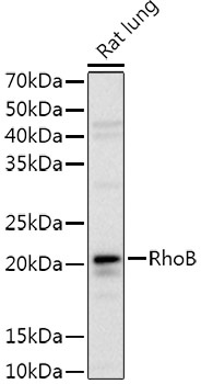
Western blot analysis of lysates from Rat lung, using RhoB Rabbit pAb (CAB2819) at 1:1000 dilution. Secondary antibody: HRP-conjugated Goat anti-Rabbit IgG (H+L) (CABS014) at 1:10000 dilution. Lysates/proteins: 25μg per lane. Blocking buffer: 3% nonfat dry milk in TBST. Detection: ECL Basic Kit (AbGn00020). Exposure time: 3s.