The RIPK1/RIP Antibody (CAB7414) is a high-quality antibody developed for reliable detection and analysis of target proteins. Raised in rabbits, this antibody demonstrates high reactivity with human samples and is validated for use in Western blot applications. By binding specifically to the RIPK1 protein, researchers can accurately detect and analyze its presence in a variety of cell types.RIPK1 is known for its critical role in regulating cell death pathways, particularly in response to stress or damage.
This antibody is validated for use in WB, IHC-P, IF/ICC, IP, ELISA applications and has demonstrated reactivity against Human, Mouse, Rat samples.
Product Name:
RIPK1/RIP Antibody
SKU:
CAB7414
Size:
20μL, 100μL
Reactivity:
Human, Mouse, Rat
Conjugate:
Unconjugated
Immunogen:
This information is considered to be commercially sensitive.
0.5μg-4μg antibody for 200μg-400μg extracts of whole cells
ELISA
Recommended starting concentration is 1 μg/mL. Please optimize the concentration based on your specific assay requirements.
Synonyms:
RIP, RIP1, AIEFL, IMD57, RIP-1, RIPK1/RIP
Positive Sample:
Raji, Jurkat treated with Etoposide, Mouse liver, C6
Cellular Localization:
Cell Membrane, Cytoplasm.
Calculated MW:
76kDa
Observed MW:
75kDa/38kDa
This gene encodes a member of the receptor-interacting protein (RIP) family of serine/threonine protein kinases. The encoded protein plays a role in inflammation and cell death in response to tissue damage, pathogen recognition, and as part of developmental regulation. RIPK1/RIPK3 kinase-mediated necrosis is referred to as necroptosis. Genetic disruption of this gene in mice results in death shortly after birth.
Purification Method
Affinity purification
Gene ID
8737
RRID
AB_2767944
Buffer Information
Store at -20℃. Avoid freeze / thaw cycles. Buffer: PBS containing 50% glycerol, preserved with proclin300 or sodium azide, pH 7.3.
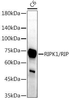
Western blot analysis of lysates from C6 cells, using RIPK1/RIP Rabbit pAb (CAB7414) at 1:1000 dilution. Secondary antibody: HRP-conjugated Goat anti-Rabbit IgG (H+L) (CABS014) at 1:10000 dilution. Lysates/proteins: 25μg per lane. Blocking buffer: 3% nonfat dry milk in TBST. Detection: ECL Enhanced Kit (AbGn00021). Exposure time: 180s.
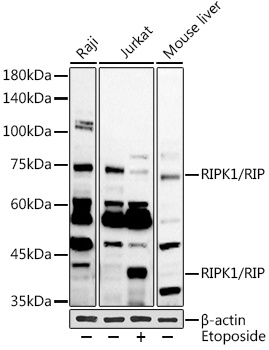
Western blot analysis of various lysates using RIPK1/RIP Rabbit pAb (CAB7414) at 1:1000 dilution. Jurkat cells were treated with Etoposide (25 uM) at 37℃ for 5 hours. Secondary antibody: HRP-conjugated Goat anti-Rabbit IgG (H+L) (CABS014) at 1:10000 dilution. Lysates/proteins: 25μg per lane. Blocking buffer: 3% nonfat dry milk in TBST. Detection: ECL Basic Kit (AbGn00020). Exposure time: 90s.
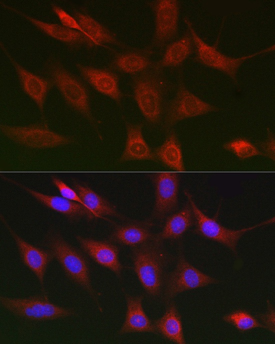
Immunofluorescence analysis of NIH/3T3 cells using RIPK1/RIP Rabbit pAb (CAB7414) at dilution of 1:100 (40x lens). Secondary antibody: Cy3-conjugated Goat anti-Rabbit IgG (H+L) (CABS007) at 1:500 dilution. Blue: DAPI for nuclear staining.