The RIPK3 Antibody (CAB5431) is a high-quality antibody developed for reliable detection and analysis of target proteins. This antibody, produced in rabbits, exhibits high specificity and sensitivity towards human samples, making it a reliable choice for Western blotting applications.RIPK3, also known as receptor-interacting protein kinase 3, plays a crucial role in mediating programmed cell death and inflammation. Dysregulation of RIPK3 has been implicated in various diseases, including inflammatory disorders and cancer.
This antibody is validated for use in WB, IHC-P, IF/ICC, ELISA applications and has demonstrated reactivity against Human, Mouse, Rat samples.
Product Name:
RIPK3 Antibody
SKU:
CAB5431
Size:
20μL, 100μL
Reactivity:
Human, Mouse, Rat
Conjugate:
Unconjugated
Immunogen:
Recombinant protein (or fragment).This information is considered to be commercially sensitive.
Recommended starting concentration is 1 μg/mL. Please optimize the concentration based on your specific assay requirements.
Synonyms:
RIP3, RIPK3
Positive Sample:
HeLa(Negative control), L929
Cellular Localization:
Cell Membrane, Cytoplasm, Mitochondrion, Cytosol.
Calculated MW:
57kDa
Observed MW:
57kDa/46-62kDa(M)
The product of this gene is a member of the receptor-interacting protein (RIP) family of serine/threonine protein kinases, and contains a C-terminal domain unique from other RIP family members. The encoded protein is predominantly localized to the cytoplasm, and can undergo nucleocytoplasmic shuttling dependent on novel nuclear localization and export signals. It is a component of the tumor necrosis factor (TNF) receptor-I signaling complex, and can induce apoptosis and weakly activate the NF-kappaB transcription factor.
Purification Method
Affinity purification
Gene ID
11035
RRID
AB_2766237
Buffer Information
Store at -20℃. Avoid freeze / thaw cycles. Buffer: PBS with 0.09% Sodium azide,50% glycerol,pH7.3.
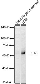
Western blot analysis of various lysates, using RIPK3 Rabbit pAb (CAB5431) at 1:3000 dilution. Secondary antibody: HRP-conjugated Goat anti-Rabbit IgG (H+L) (CABS014) at 1:10000 dilution. Lysates/proteins: 25μg per lane. Blocking buffer: 3% nonfat dry milk in TBST. Detection: ECL Basic Kit (AbGn00020). Exposure time: 30s.
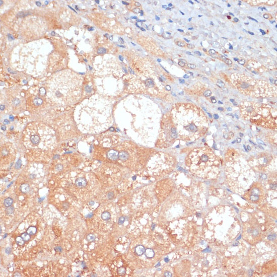
Immunohistochemistry analysis of paraffin-embedded Human liver cancer using RIPK3 Rabbit pAb (CAB5431) at dilution of 1:100 (40x lens). Microwave antigen retrieval performed with 0.01M PBS Buffer (pH 7.2) prior to IHC staining.
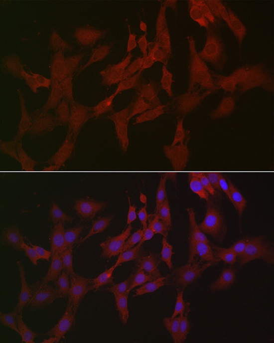
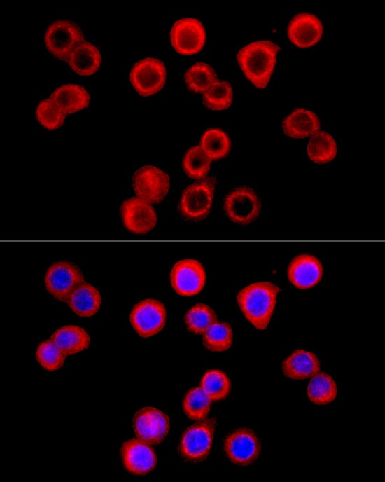
Immunofluorescence analysis of C6 cells using RIPK3 Rabbit pAb (CAB5431) at dilution of 1:50 (40x lens). Secondary antibody: Cy3-conjugated Goat anti-Rabbit IgG (H+L) (CABS007) at 1:500 dilution. Blue: DAPI for nuclear staining.
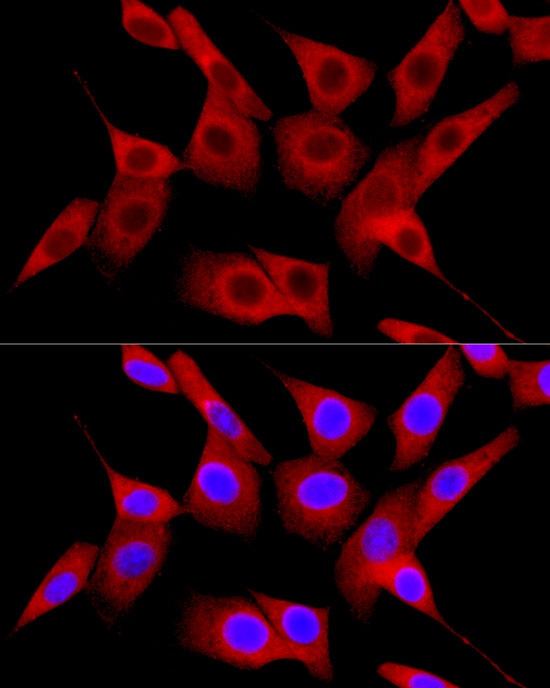
Immunofluorescence analysis of NIH/3T3 cells using RIPK3 Rabbit pAb (CAB5431) at dilution of 1:50 (40x lens). Secondary antibody: Cy3-conjugated Goat anti-Rabbit IgG (H+L) (CABS007) at 1:500 dilution. Blue: DAPI for nuclear staining.
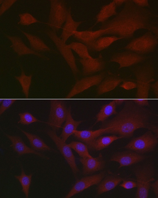
Immunofluorescence analysis of HT-29 cells using RIPK3 Rabbit pAb (CAB5431) at dilution of 1:100 (40x lens). Secondary antibody: Cy3-conjugated Goat anti-Rabbit IgG (H+L) (CABS007) at 1:500 dilution. Blue: DAPI for nuclear staining.
Immunofluorescence analysis of NIH/3T3 cells using RIPK3 Rabbit pAb (CAB5431) at dilution of 1:100 (40x lens). Secondary antibody: Cy3-conjugated Goat anti-Rabbit IgG (H+L) (CABS007) at 1:500 dilution. Blue: DAPI for nuclear staining.