The RNF126 Antibody (CAB20015) is a high-quality antibody developed for reliable detection and analysis of target proteins. This antibody, raised in rabbits, exhibits high reactivity with human samples and has been validated for use in Western blot applications. By binding to the RNF126 protein, this antibody allows for efficient detection and analysis in a variety of cell types, making it an essential tool for studies in cell biology and protein degradation pathways. RNF126 is a critical player in the regulation of protein turnover and has been linked to various cellular processes, including cell cycle progression, DNA repair, and signaling pathways.
This antibody is validated for use in WB, IHC-P, IF/ICC, ELISA applications and has demonstrated reactivity against Human, Mouse, Rat samples.
Product Name:
RNF126 Antibody
SKU:
CAB20015
Size:
20μL, 100μL
Reactivity:
Human, Mouse, Rat
Conjugate:
Unconjugated
Immunogen:
Synthetic peptide. This information is considered to be commercially sensitive.
Recommended starting concentration is 1 μg/mL. Please optimize the concentration based on your specific assay requirements.
Synonyms:
RNF126
Positive Sample:
293T, Rat testis
Cellular Localization:
Cytoplasm, Nucleus.
Calculated MW:
34kDa
Observed MW:
36-38kDa
The protein encoded by this gene contains a RING finger domain, a motif present in a variety of functionally distinct proteins and known to be involved in protein-protein and protein-DNA interactions.
Purification Method
Affinity purification
Gene ID
55658
RRID
AB_2862919
Buffer Information
Store at -20℃. Avoid freeze / thaw cycles. Buffer: PBS containing 50% glycerol, preserved with proclin300 or sodium azide, pH 7.3.
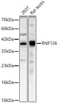
Western blot analysis of various lysates, using RNF126 Rabbit pAb (CAB20015) at 1:2000 dilution. Secondary antibody: HRP-conjugated Goat anti-Rabbit IgG (H+L) (CABS014) at 1:10000 dilution. Lysates/proteins: 25μg per lane. Blocking buffer: 3% nonfat dry milk in TBST. Detection: ECL Basic Kit (AbGn00020). Exposure time: 30s.
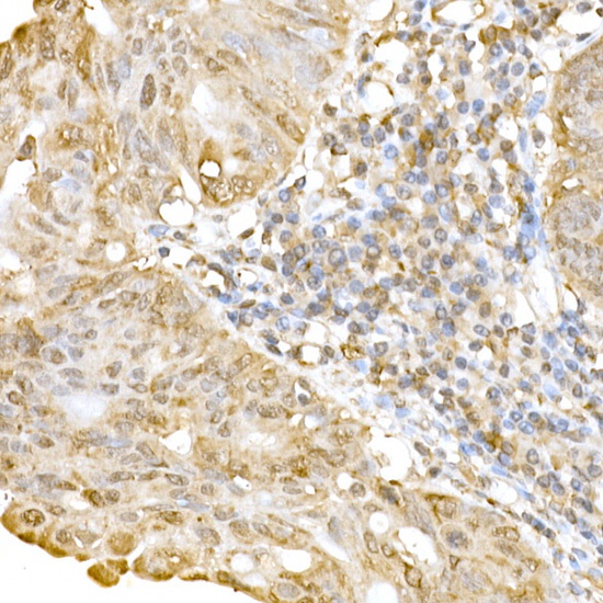
Immunohistochemistry analysis of paraffin-embedded Human colon carcinoma using RNF126 Rabbit pAb (CAB20015) at dilution of 1:250 (40x lens). High pressure antigen retrieval performed with 0.01M Citrate buffer (pH 6.0) prior to IHC staining.
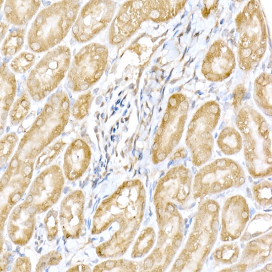
Immunohistochemistry analysis of paraffin-embedded Mouse kidney using RNF126 Rabbit pAb (CAB20015) at dilution of 1:250 (40x lens). High pressure antigen retrieval performed with 0.01M Citrate buffer (pH 6.0) prior to IHC staining.
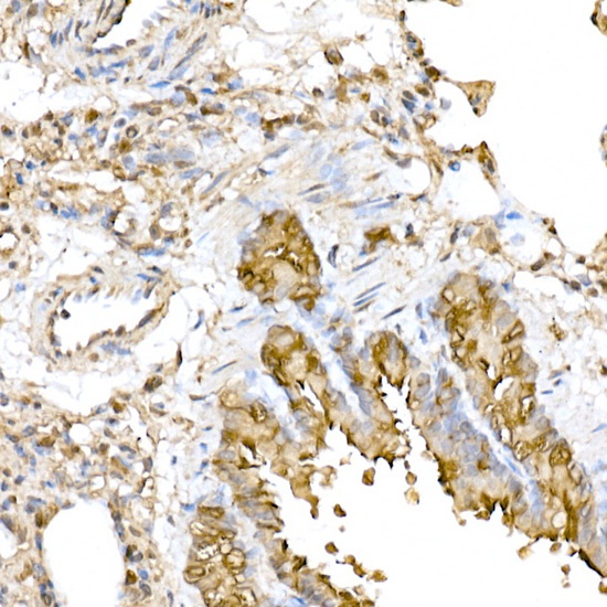
Immunohistochemistry analysis of paraffin-embedded Rat lung using RNF126 Rabbit pAb (CAB20015) at dilution of 1:250 (40x lens). High pressure antigen retrieval performed with 0.01M Citrate buffer (pH 6.0) prior to IHC staining.
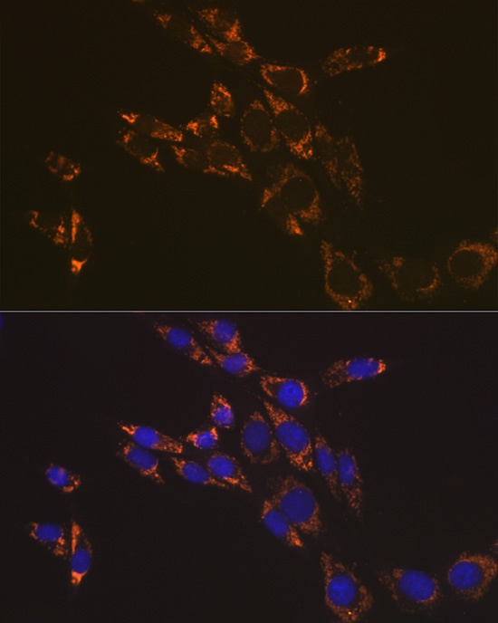
Immunofluorescence analysis of NIH-3T3 cells using RNF126 Rabbit pAb (CAB20015) at dilution of 1:100 (40x lens). Secondary antibody: Cy3-conjugated Goat anti-Rabbit IgG (H+L) (CABS007) at 1:500 dilution. Blue: DAPI for nuclear staining.