The RNH1 Antibody (CAB4079) is a high-quality antibody developed for reliable detection and analysis of target proteins. This high-quality antibody is produced in rabbits and exhibits strong reactivity with human samples, making it ideal for a variety of research applications.RNH1 is known to play a crucial role in maintaining RNA integrity and stability within the cell. Dysregulation of RNH1 has been linked to various diseases, including cancer and neurodegenerative disorders. By using the RNH1 Polyclonal Antibody, researchers can accurately detect and analyze the expression of RNH1 in different cell types and tissues.
This antibody is validated for use in WB, IF/ICC, ELISA applications and has demonstrated reactivity against Human, Mouse, Rat samples.
Product Name:
RNH1 Antibody
SKU:
CAB4079
Size:
20μL, 100μL
Reactivity:
Human, Mouse, Rat
Conjugate:
Unconjugated
Immunogen:
Recombinant protein (or fragment).This information is considered to be commercially sensitive.
Recommended starting concentration is 1 μg/mL. Please optimize the concentration based on your specific assay requirements.
Synonyms:
RAI, RNH, RNH1
Positive Sample:
Raji, U-87MG, HT-29, HeLa, Mouse liver, Mouse kidney, Mouse lung, Rat brain, Rat lung
Cellular Localization:
Cytoplasm.
Calculated MW:
50kDa
Observed MW:
43kDa
Placental ribonuclease inhibitor (PRI) is a member of a family of proteinaceous cytoplasmic RNase inhibitors that occur in many tissues and bind to both intracellular and extracellular RNases (summarized by Lee et al., 1988 [PubMed 3219362]). In addition to control of intracellular RNases, the inhibitor may have a role in the regulation of angiogenin (MIM 105850). Ribonuclease inhibitor, of 50,000 Da, binds to ribonucleases and holds them in a latent form. Since neutral and alkaline ribonucleases probably play a critical role in the turnover of RNA in eukaryotic cells, RNH may be essential for control of mRNA turnover; the interaction of eukaryotic cells with ribonuclease may be reversible in vivo.
Purification Method
Affinity purification
Gene ID
6050
RRID
AB_2765491
Buffer Information
Store at -20℃. Avoid freeze / thaw cycles. Buffer: PBS containing 50% glycerol, preserved with proclin300 or sodium azide, pH 7.3.
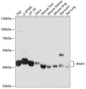
Western blot analysis of various lysates using RNH1 Rabbit pAb (CAB4079) at 1:1000 dilution. Secondary antibody: HRP-conjugated Goat anti-Rabbit IgG (H+L) (CABS014) at 1:10000 dilution. Lysates/proteins: 25μg per lane. Blocking buffer: 3% nonfat dry milk in TBST. Detection: ECL Basic Kit (AbGn00020). Exposure time: 1s.
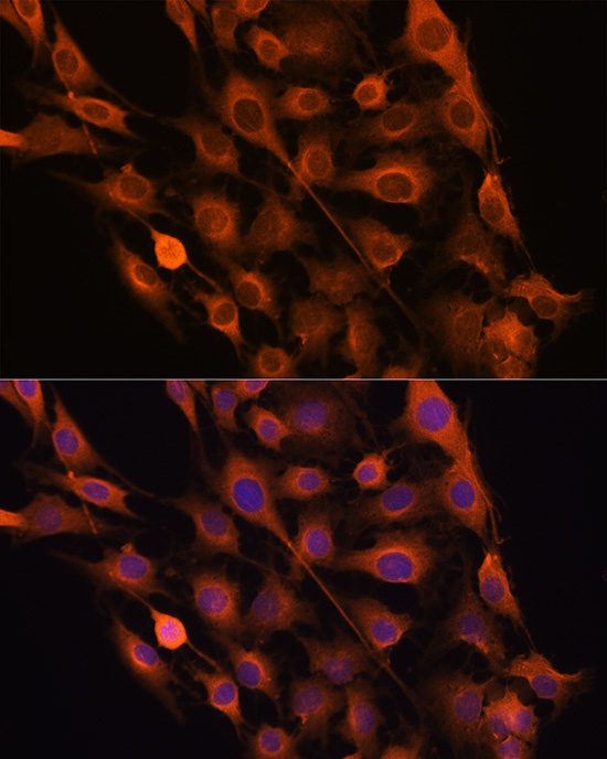
Immunofluorescence analysis of C6 cells using RNH1 Rabbit pAb (CAB4079) at dilution of 100 (40x lens). Secondary antibody: Cy3-conjugated Goat anti-Rabbit IgG (H+L) (CABS007) at 1:500 dilution. Blue: DAPI for nuclear staining.
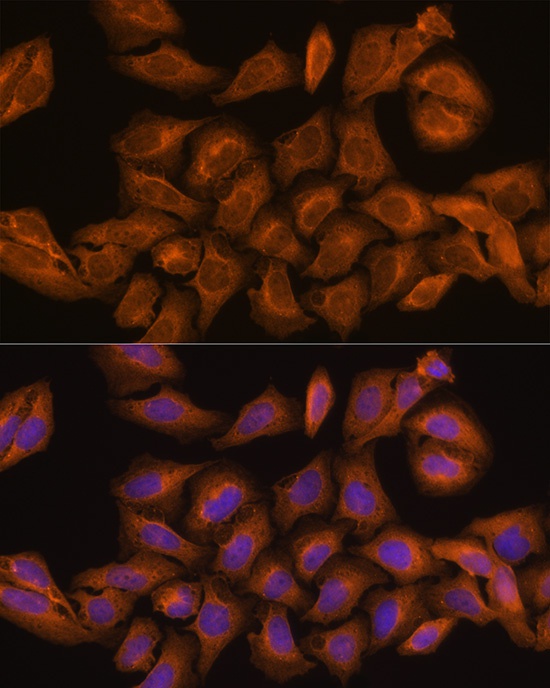
Immunofluorescence analysis of U-2 OS cells using RNH1 Rabbit pAb (CAB4079) at dilution of 100 (40x lens). Secondary antibody: Cy3-conjugated Goat anti-Rabbit IgG (H+L) (CABS007) at 1:500 dilution. Blue: DAPI for nuclear staining.