The ROCK2 Antibody (CAB5698) is a high-quality antibody developed for reliable detection and analysis of target proteins. This antibody, produced in rabbits, shows high reactivity with human samples and has been validated for use in Western blot applications. By binding specifically to the ROCK2 protein, this antibody enables accurate detection and analysis in a variety of cell types, making it ideal for investigations in cell biology, cancer research, and drug development.ROCK2, a member of the Rho-associated coiled-coil containing protein kinase family, plays a crucial role in various cellular processes including cell adhesion, motility, and cell division.
This antibody is validated for use in WB, IHC-P, IF/ICC, ELISA applications and has demonstrated reactivity against Human, Mouse, Rat samples.
Product Name:
ROCK2 Antibody
SKU:
CAB5698
Size:
20μL, 100μL
Reactivity:
Human, Mouse, Rat
Conjugate:
Unconjugated
Immunogen:
Recombinant protein (or fragment).This information is considered to be commercially sensitive.
The protein encoded by this gene is a serine/threonine kinase that regulates cytokinesis, smooth muscle contraction, the formation of actin stress fibers and focal adhesions, and the activation of the c-fos serum response element. This protein, which is an isozyme of ROCK1 is a target for the small GTPase Rho.
Purification Method
Affinity purification
Gene ID
9475
RRID
AB_2766457
Buffer Information
Store at -20℃. Avoid freeze / thaw cycles. Buffer: PBS containing 50% glycerol, preserved with proclin300 or sodium azide, pH 7.3.
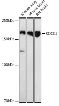
Western blot analysis of various lysates using ROCK2 Rabbit pAb (CAB5698) at 1:1000 dilution. Secondary antibody: HRP-conjugated Goat anti-Rabbit IgG (H+L) (CABS014) at 1:10000 dilution. Lysates/proteins: 25μg per lane. Blocking buffer: 3% nonfat dry milk in TBST. Detection: ECL Basic Kit (AbGn00020). Exposure time: 180s.
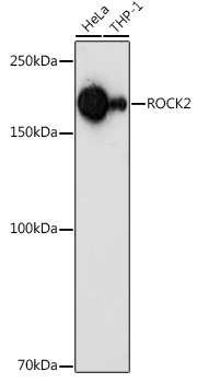
Western blot analysis of various lysates using ROCK2 Rabbit pAb (CAB5698) at 1:1000 dilution. Secondary antibody: HRP-conjugated Goat anti-Rabbit IgG (H+L) (CABS014) at 1:10000 dilution. Lysates/proteins: 25μg per lane. Blocking buffer: 3% nonfat dry milk in TBST. Detection: ECL Basic Kit (AbGn00020). Exposure time: 180s.
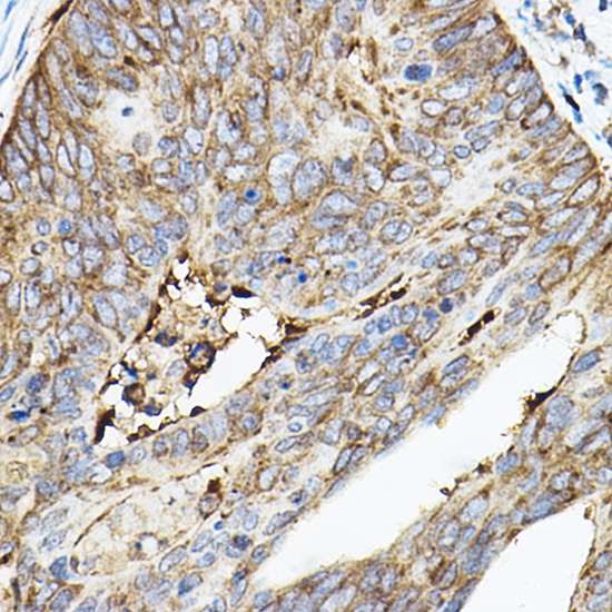
Immunohistochemistry analysis of paraffin-embedded Human colon carcinoma using ROCK2 Rabbit pAb (CAB5698) at dilution of 1:50 (40x lens). High pressure antigen retrieval performed with 0.01M Citrate buffer (pH 6.0) prior to IHC staining.
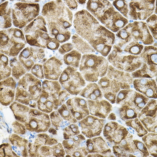
Immunohistochemistry analysis of paraffin-embedded Mouse pancreas using ROCK2 Rabbit pAb (CAB5698) at dilution of 1:50 (40x lens). High pressure antigen retrieval performed with 0.01M Citrate buffer (pH 6.0) prior to IHC staining.
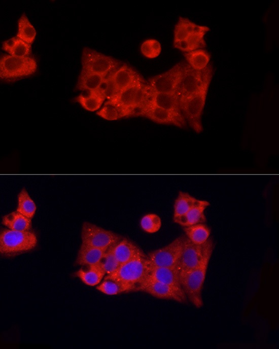
Immunofluorescence analysis of HepG2 cells using ROCK2 Rabbit pAb (CAB5698) at dilution of 1:100 (40x lens). Secondary antibody: Cy3-conjugated Goat anti-Rabbit IgG (H+L) (CABS007) at 1:500 dilution. Blue: DAPI for nuclear staining.
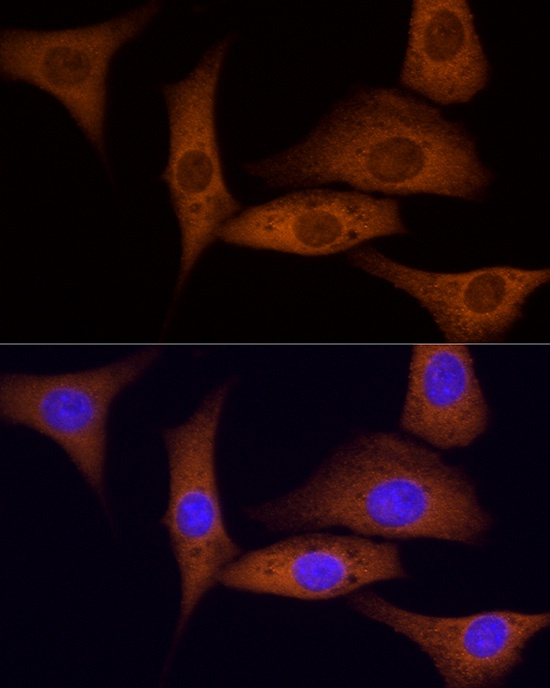
Immunofluorescence analysis of NIH/3T3 cells using ROCK2 Rabbit pAb (CAB5698) at dilution of 1:100 (40x lens). Secondary antibody: Cy3-conjugated Goat anti-Rabbit IgG (H+L) (CABS007) at 1:500 dilution. Blue: DAPI for nuclear staining.
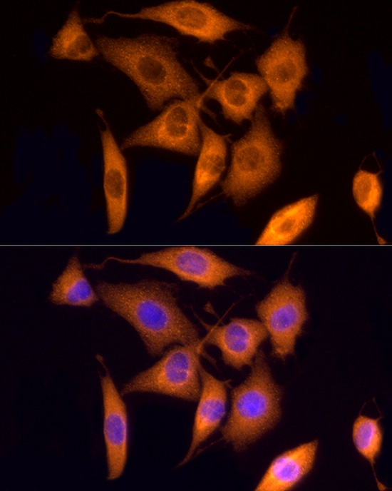
Immunofluorescence analysis of PC-12 cells using ROCK2 Rabbit pAb (CAB5698) at dilution of 1:100 (40x lens). Secondary antibody: Cy3-conjugated Goat anti-Rabbit IgG (H+L) (CABS007) at 1:500 dilution. Blue: DAPI for nuclear staining.


")


