The RRM2 Antibody (CAB5255) is a high-quality antibody developed for reliable detection and analysis of target proteins. This antibody, produced in rabbits, is highly specific to human samples and has been validated for use in Western blot applications. By binding to the RRM2 protein, researchers can accurately detect and analyze its expression in various cell types, making it an essential tool for studies in molecular biology and cancer research.
This antibody is validated for use in WB, IHC-P, IF/ICC, IP, ELISA applications and has demonstrated reactivity against Human, Mouse, Rat samples.
Product Name:
RRM2 Antibody
SKU:
CAB5255
Size:
20μL, 100μL
Reactivity:
Human, Mouse, Rat
Conjugate:
Unconjugated
Immunogen:
Recombinant protein (or fragment).This information is considered to be commercially sensitive.
0.5μg-4μg antibody for 200μg-400μg extracts of whole cells
ELISA
Recommended starting concentration is 1 μg/mL. Please optimize the concentration based on your specific assay requirements.
Synonyms:
R2, RR2, RR2M, C2orf48, RRM2
Positive Sample:
HeLa
Cellular Localization:
Cytoplasm.
Calculated MW:
45kDa
Observed MW:
45kDa
This gene encodes one of two non-identical subunits for ribonucleotide reductase. This reductase catalyzes the formation of deoxyribonucleotides from ribonucleotides. Synthesis of the encoded protein (M2) is regulated in a cell-cycle dependent fashion. Transcription from this gene can initiate from alternative promoters, which results in two isoforms that differ in the lengths of their N-termini. Related pseudogenes have been identified on chromosomes 1 and X.
Purification Method
Affinity purification
Gene ID
6241
RRID
AB_2766082
Buffer Information
Store at -20℃. Avoid freeze / thaw cycles. Buffer: PBS containing 50% glycerol, preserved with proclin300 or sodium azide, pH 7.3.
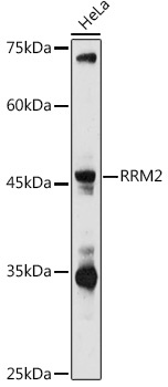
Western blot analysis of lysates from HeLa cells, using RAbGn2 Rabbit pAb (CAB5255) at 1:1000 dilution. Secondary antibody: HRP-conjugated Goat anti-Rabbit IgG (H+L) (CABS014) at 1:10000 dilution. Lysates/proteins: 25μg per lane. Blocking buffer: 3% nonfat dry milk in TBST. Detection: ECL Basic Kit (AbGn00020). Exposure time: 180s.
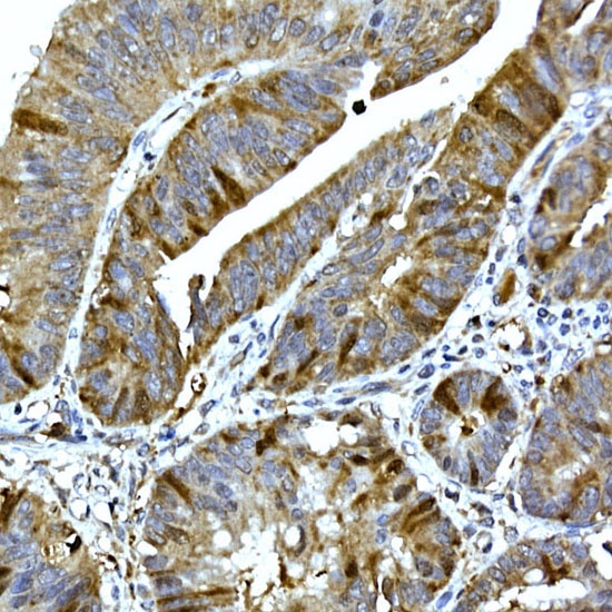
Immunohistochemistry analysis of paraffin-embedded Human colon carcinoma using RAbGn2 Rabbit pAb (CAB5255) at dilution of 1:100 (40x lens). High pressure antigen retrieval performed with 0.01M Citrate buffer (pH 6.0) prior to IHC staining.
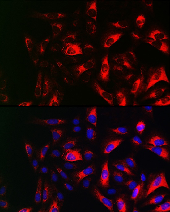
Immunofluorescence analysis of U2OS cells using RAbGn2 Rabbit pAb (CAB5255) at dilution of 1:100 (40x lens). Secondary antibody: Cy3-conjugated Goat anti-Rabbit IgG (H+L) (CABS007) at 1:500 dilution. Blue: DAPI for nuclear staining.
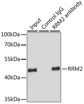
Immunoprecipitation analysis of 200 μg extracts of HeLa cells using 1 μg RAbGn2 antibody (CAB5255). Western blot was performed from the immunoprecipitate using RAbGn2 antibody (CAB5255) at a dilution of 1:1000.


")


