The SCFD1 Antibody (CAB8835) is a high-quality antibody developed for reliable detection and analysis of target proteins. This antibody, produced in rabbits, demonstrates high reactivity with human samples and is specifically validated for use in Western blot applications.SCFD1, a key player in cellular processes, is known for its role in mediating protein degradation and regulating protein turnover. Its involvement in these pathways makes it a relevant target for research in the fields of cell biology and signal transduction. By utilizing the SCFD1 Polyclonal Antibody, researchers can effectively detect and analyze SCFD1 protein levels in various cell types, providing valuable insights into its function and potential therapeutic applications.
This antibody is validated for use in WB, IHC-P, ELISA applications and has demonstrated reactivity against Human, Mouse, Rat samples.
Product Name:
SCFD1 Antibody
SKU:
CAB8835
Size:
20μL, 100μL
Reactivity:
Human, Mouse, Rat
Conjugate:
Unconjugated
Immunogen:
Recombinant protein (or fragment).This information is considered to be commercially sensitive.
Predicted to enable syntaxin binding activity. Involved in negative regulation of autophagosome assembly; regulation of protein transport; and response to toxic substance. Located in cis-Golgi network.
Purification Method
Affinity purification
Gene ID
23256
RRID
AB_2772151
Buffer Information
Store at -20℃. Avoid freeze / thaw cycles. Buffer: PBS containing 50% glycerol, preserved with proclin300 or sodium azide, pH 7.3.
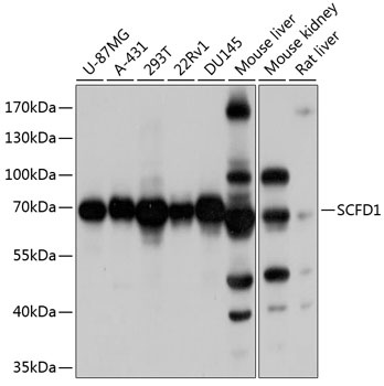
Western blot analysis of various lysates using SCFD1 Rabbit pAb (CAB8835) at 1:1000 dilution. Secondary antibody: HRP-conjugated Goat anti-Rabbit IgG (H+L) (CABS014) at 1:10000 dilution. Lysates/proteins: 25μg per lane. Blocking buffer: 3% nonfat dry milk in TBST. Detection: ECL Basic Kit (AbGn00020). Exposure time: 1s.
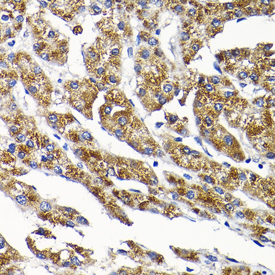
Immunohistochemistry analysis of paraffin-embedded Human liver cancer using SCFD1 Rabbit pAb (CAB8835) at dilution of 1:200 (40x lens). High pressure antigen retrieval performed with 0.01M Citrate buffer (pH 6.0) prior to IHC staining.
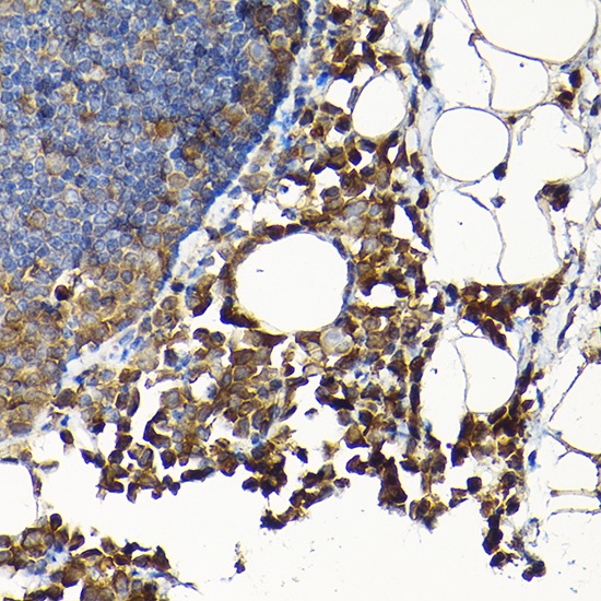
Immunohistochemistry analysis of paraffin-embedded Rat lung using SCFD1 Rabbit pAb (CAB8835) at dilution of 1:200 (40x lens). High pressure antigen retrieval performed with 0.01M Citrate buffer (pH 6.0) prior to IHC staining.