The SDHA Monoclonal Antibody (CAB13852) is a high-quality antibody developed for reliable detection and analysis of target proteins. This gene encodes a major catalytic subunit of succinate-ubiquinone oxidoreductase, a complex of the mitochondrial respiratory chain. The complex is composed of four nuclear-encoded subunits and is localized in the mitochondrial inner membrane. Mutations in this gene have been associated with a form of mitochondrial respiratory chain deficiency known as Leigh Syndrome. A pseudogene has been identified on chromosome 3q29. Alternatively spliced transcript variants encoding different isoforms have been found for this gene. RRID AB_2861697 Gene ID 6389 Swiss Prot Synonym FP; PGL5; SDH1; SDH2; SDHF; CMD1GG; MC2DN1; NDAXOA; SDHA
This antibody is validated for use in WB, IF/ICC, ELISA, IF-P applications and has demonstrated reactivity against Human, Mouse, Rat samples.
Product Name:
SDHA Monoclonal Antibody
SKU:
CAB13852
Size:
100μL, 20μL
Reactivity:
Human, Mouse, Rat
Clone Number:
ARC0726
Conjugate:
Unconjugated
Immunogen:
Synthetic peptide. This information is considered to be commercially sensitive.
Tested Applications:
WBIF/ICCELISAIF-P
Recommended Dilution:
WB
1:1000 - 1:6000
IF
/
ICC
1:200 - 1:800
IF-P
1:200 - 1:800
ELISA
Recommended starting concentration is 1 μg/mL. Please optimize the concentration based on your specific assay requirements.
This gene encodes a major catalytic subunit of succinate-ubiquinone oxidoreductase, a complex of the mitochondrial respiratory chain. The complex is composed of four nuclear-encoded subunits and is localized in the mitochondrial inner membrane. Mutations in this gene have been associated with a form of mitochondrial respiratory chain deficiency known as Leigh Syndrome. A pseudogene has been identified on chromosome 3q29. Alternatively spliced transcript variants encoding different isoforms have been found for this gene. RRID AB_2861697 Gene ID 6389 Swiss Prot Synonym FP; PGL5; SDH1; SDH2; SDHF; CMD1GG; MC2DN1; NDAXOA; SDHA
Purification Method:
Affinity purification
Gene ID:
6389
RRID:
AB_2861697
Buffer Information:
Store at -20℃. Avoid freeze / thaw cycles. Buffer: PBS containing 50% glycerol and 0.05% BSA, preserved with proclin300 or sodium azide, pH 7.3.
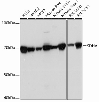
Western blot analysis of various lysates using SDHA Rabbit mAb (CAB13852) at 1:1000 dilution incubated overnight at 4℃. Secondary antibody: HRP-conjugated Goat anti-Rabbit IgG (H+L) (AS014) at 1:10000 dilution. Lysates/proteins: 25 μg per lane. Blocking buffer: 3% nonfat dry milk in TBST. Detection: ECL Basic Kit (AbGn00020). Exposure time: 1 s.
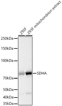
Western blot analysis of lysates from 293F cells using SDHA Rabbit mAb (CAB13852) at 1:1000 dilution incubated overnight at 4℃. Mitochondrion extracts derived from 293F cells. Secondary antibody: HRP-conjugated Goat anti-Rabbit IgG (H+L) (AS014) at 1:10000 dilution. Lysates/proteins: 25 μg per lane. Blocking buffer: 3% nonfat dry milk in TBST. Detection: ECL Basic Kit (AbGn00020). Exposure time: 1 s.
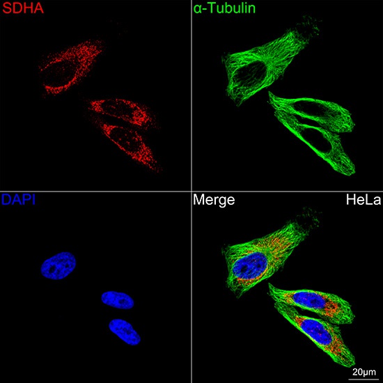
Confocal imaging of HeLa cells using SDHA Rabbit mAb (CAB13852, dilution 1:200) followed by a further incubation with Cy3-conjugated Goat anti-Rabbit IgG (H+L) (AS007, dilution 1:500) (Red). The cells were counterstained with α-Tubulin Mouse mAb (AC012, dilution 1:400) followed by incubation with ABflo® 488-conjugated Goat Anti-Mouse IgG (H+L) Ab (AS076, dilution 1:500) (Green). DAPI was used for nuclear staining (Blue). Objective: 100x.