The Septin 1 Antibody (CAB17471) is a high-quality antibody developed for reliable detection and analysis of target proteins. This gene is a member of the septin family of GTPases. Members of this family are required for cytokinesis and the maintenance of cellular morphology. This gene encodes a protein that can form homo- and heterooligomeric filaments, and may contribute to the formation of neurofibrillary tangles in Alzheimer's disease. Alternatively spliced transcript variants have been found but the full-length nature of these variants has not been determined.
This antibody is validated for use in WB, IHC-P, ELISA applications and has demonstrated reactivity against Human, Mouse, Rat samples.
Product Name:
Septin 1 Antibody
SKU:
CAB17471
Size:
100μL, 20μL
Reactivity:
Human, Mouse, Rat
Conjugate:
Unconjugated
Immunogen:
Recombinant protein (or fragment).This information is considered to be commercially sensitive.
Tested Applications:
WBIHC-PELISA
Recommended Dilution:
WB
1:500 - 1:1000
IHC-P
1:50 - 1:100
ELISA
Recommended starting concentration is 1 μg/mL. Please optimize the concentration based on your specific assay requirements.
This gene is a member of the septin family of GTPases. Members of this family are required for cytokinesis and the maintenance of cellular morphology. This gene encodes a protein that can form homo- and heterooligomeric filaments, and may contribute to the formation of neurofibrillary tangles in Alzheimer's disease. Alternatively spliced transcript variants have been found but the full-length nature of these variants has not been determined.
Purification Method
Affinity purification
Gene ID
1731
RRID
AB_2772179
Buffer Information
Store at -20℃. Avoid freeze / thaw cycles. Buffer: PBS with 0.01% thimerosal,50% glycerol,pH7.3.
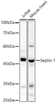
Western blot analysis of various lysates, using Septin 1 Rabbit pAb (CAB17471) at 1:1000 dilution. Secondary antibody: HRP-conjugated Goat anti-Rabbit IgG (H+L) (AS014) at 1:10000 dilution. Lysates/proteins: 25μg per lane. Blocking buffer: 3% nonfat dry milk in TBST. Detection: ECL Basic Kit (AbGn00020). Exposure time: 20s.
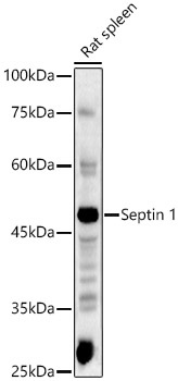
Western blot analysis of lysates from Rat spleen, using Septin 1 Rabbit pAb (CAB17471) at 1:1000 dilution. Secondary antibody: HRP-conjugated Goat anti-Rabbit IgG (H+L) (AS014) at 1:10000 dilution. Lysates/proteins: 25μg per lane. Blocking buffer: 3% nonfat dry milk in TBST. Detection: ECL Basic Kit (AbGn00020). Exposure time: 180s.
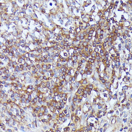
Immunohistochemistry analysis of paraffin-embedded Human appendix using Septin 1 Rabbit pAb (CAB17471) at dilution of 1:100 (40x lens). Microwave antigen retrieval performed with 0.01M PBS Buffer (pH 7.2) prior to IHC staining.
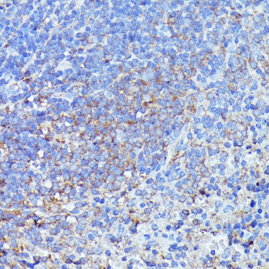
Immunohistochemistry analysis of paraffin-embedded Mouse spleen using Septin 1 Rabbit pAb (CAB17471) at dilution of 1:100 (40x lens). Microwave antigen retrieval performed with 0.01M PBS Buffer (pH 7.2) prior to IHC staining.