The SHC1 Antibody (CAB7725) is a high-quality antibody developed for reliable detection and analysis of target proteins. This antibody, produced in rabbits, has high reactivity with human samples and has been validated for use in Western blot applications. By specifically binding to the SHC1 protein, this antibody allows for reliable detection and analysis in various cell types, making it ideal for investigations in molecular biology, cell signaling, and cancer research.SHC1 is a key adaptor protein that plays a pivotal role in transmitting extracellular signals to the cell interior, influencing processes such as cell growth, differentiation, and survival.
This antibody is validated for use in WB, IHC-P, IF/ICC, ELISA applications and has demonstrated reactivity against Human, Mouse, Rat samples.
Product Name:
SHC1 Antibody
SKU:
CAB7725
Size:
20μL, 100μL
Reactivity:
Human, Mouse, Rat
Conjugate:
Unconjugated
Immunogen:
Synthetic peptide. This information is considered to be commercially sensitive.
Recommended starting concentration is 1 μg/mL. Please optimize the concentration based on your specific assay requirements.
Synonyms:
SHC, SHCA, SHC1
Positive Sample:
Jurkat, C6, Mouse lung, Mouse spleen, Rat liver
Cellular Localization:
Cytoplasm, Mitochondrion, Mitochondrion Matrix.
Calculated MW:
63kDa
Observed MW:
66kDa
This gene encodes three main isoforms that differ in activities and subcellular location. While all three are adapter proteins in signal transduction pathways, the longest (p66Shc) may be involved in regulating life span and the effects of reactive oxygen species. The other two isoforms, p52Shc and p46Shc, link activated receptor tyrosine kinases to the Ras pathway by recruitment of the GRB2/SOS complex. p66Shc is not involved in Ras activation. Unlike the other two isoforms, p46Shc is targeted to the mitochondrial matrix. Several transcript variants encoding different isoforms have been found for this gene.
Purification Method
Affinity purification
Gene ID
6464
RRID
AB_2772210
Buffer Information
Store at -20℃. Avoid freeze / thaw cycles. Buffer: PBS containing 50% glycerol, preserved with proclin300 or sodium azide, pH 7.3.
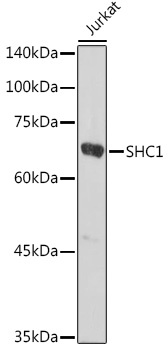
Western blot analysis of lysates from Jurkat cells, using SHC1 Rabbit pAb (CAB7725) at 1:1000 dilution. Secondary antibody: HRP-conjugated Goat anti-Rabbit IgG (H+L) (CABS014) at 1:10000 dilution. Lysates/proteins: 25μg per lane. Blocking buffer: 3% nonfat dry milk in TBST. Detection: ECL Basic Kit (AbGn00020). Exposure time: 30s.
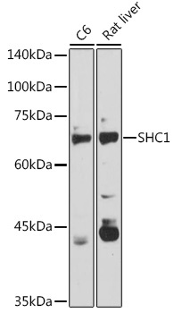
Western blot analysis of various lysates using SHC1 Rabbit pAb (CAB7725) at 1:1000 dilution. Secondary antibody: HRP-conjugated Goat anti-Rabbit IgG (H+L) (CABS014) at 1:10000 dilution. Lysates/proteins: 25μg per lane. Blocking buffer: 3% nonfat dry milk in TBST. Detection: ECL Basic Kit (AbGn00020). Exposure time: 90s.
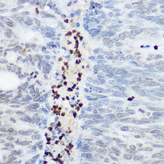
Immunohistochemistry analysis of paraffin-embedded Human colon carcinoma using SHC1 Rabbit pAb (CAB7725) at dilution of 1:100 (40x lens). High pressure antigen retrieval performed with 0.01M Citrate buffer (pH 6.0) prior to IHC staining.
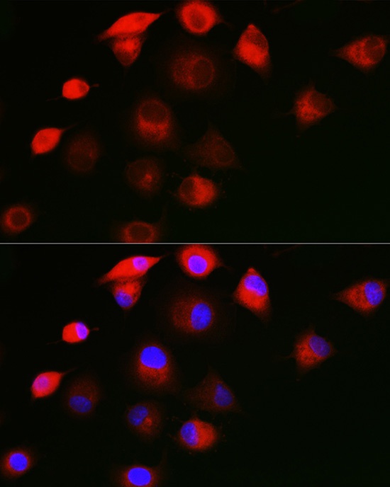
Immunofluorescence analysis of A549 cells using SHC1 Rabbit pAb (CAB7725) at dilution of 1:50 (40x lens). Secondary antibody: Cy3-conjugated Goat anti-Rabbit IgG (H+L) (CABS007) at 1:500 dilution. Blue: DAPI for nuclear staining.